Preclinical Bone & Body Composition Imaging
iNSiGHT DXA
Preclinical in vivo DXA/DEXA for quantitative bone and body-composition analysis in small-animal studies
Measure BMD, BMC, fat mass, lean mass, and related body-composition endpoints from a 25-second scan. Low-dose X-ray acquisition supports repeated longitudinal measurements in small-animal studies.
iNSiGHT DXA Resources
iNSiGHT DXA System Overview

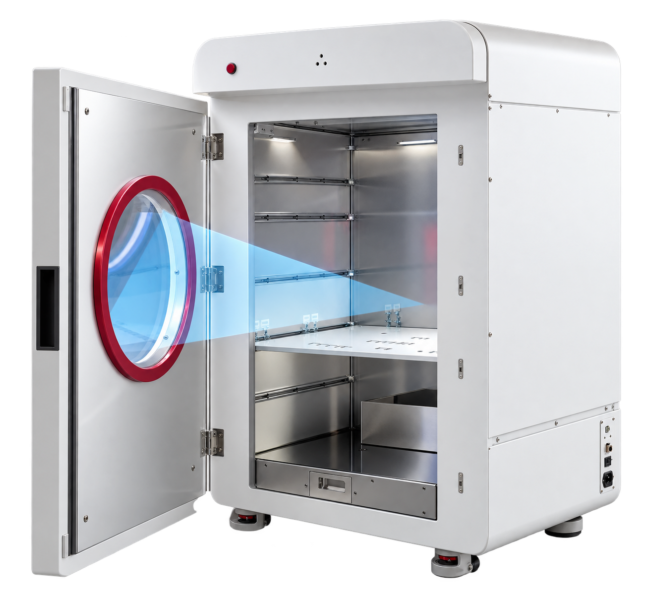
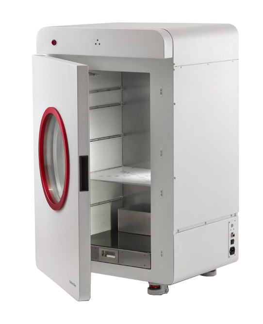
The iNSiGHT DXA system is a fully shielded preclinical dual-energy X-ray absorptiometry (DXA/DEXA) platform for quantitative assessment of bone mineral and body-composition endpoints in small-animal studies. It measures bone mineral density (BMD), bone mineral content (BMC), fat mass, lean mass, and related body-composition parameters.
With a 25-second acquisition and low-dose X-ray imaging, iNSiGHT supports repeated longitudinal measurements. Routine in vivo acquisition uses anesthesia, and measurements can be evaluated across the whole animal or within user-defined regions of interest.
Key Imaging & Workflow Benefits
From fast, low-dose acquisition to quantitative bone and body-composition analysis, iNSiGHT DXA is designed to support efficient longitudinal preclinical imaging workflows.
DXA Acquisition & Quantitative Analysis
iNSiGHT uses dual-energy X-rays to classify tissue into fat mass, non-bone/lean mass, and bone mineral content. From a single 25-second scan, the system generates quantitative bone and body-composition measurements for the whole animal or user-defined regions of interest.
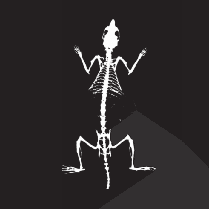
Bone Measurements
Bone mineral density, bone mineral content, bone area, and specific bone-length measurements drawn from the 2D X-ray image.
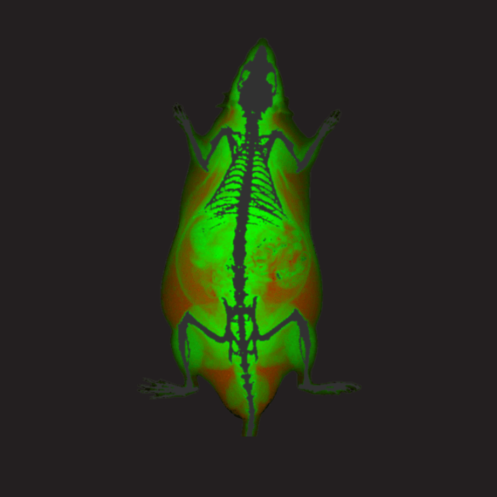
Body Composition
Quantifies fat tissue mass and percentage, lean/non-bone tissue mass and percentage, tissue area, and total mass.
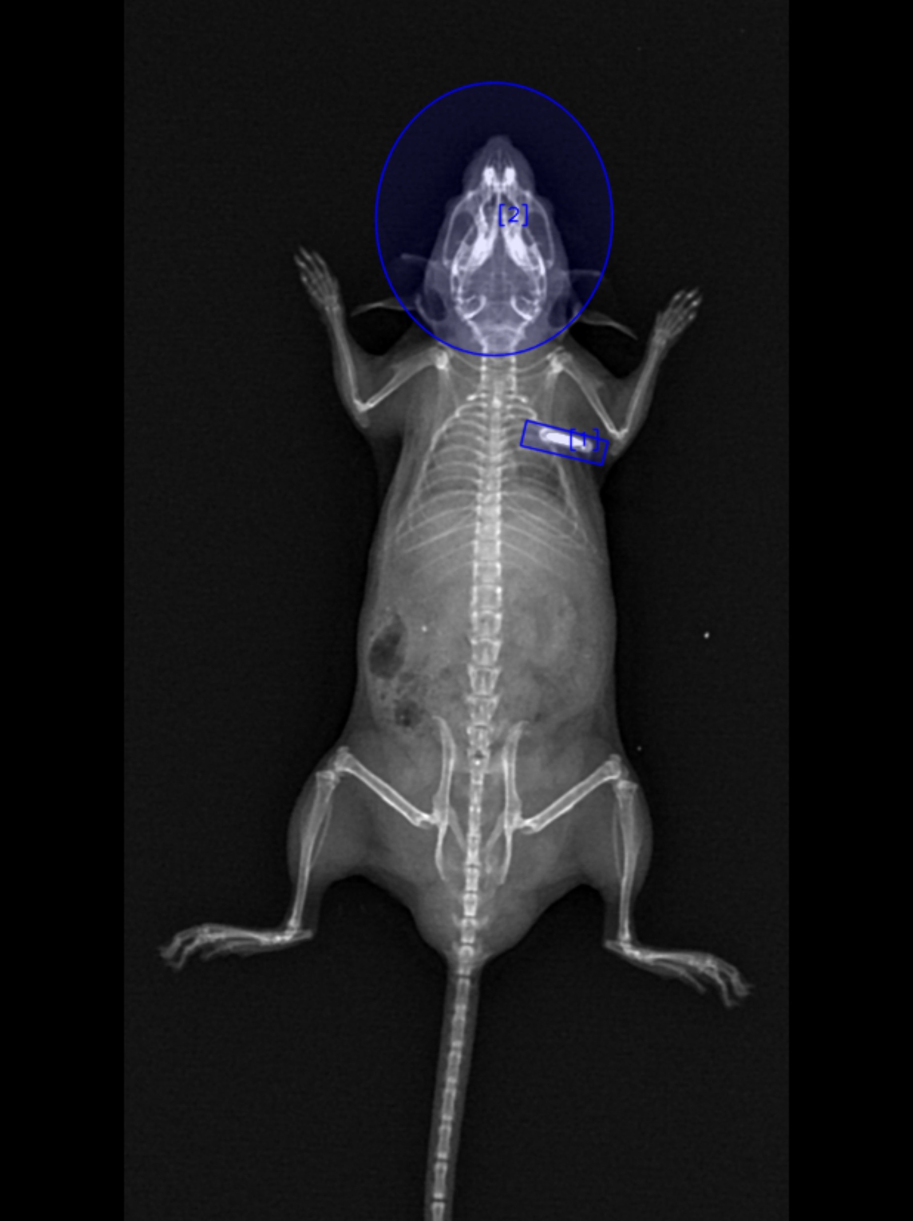
Whole-Animal & ROI Analysis
Measurements can be calculated from the whole animal or from user-defined regions of interest.
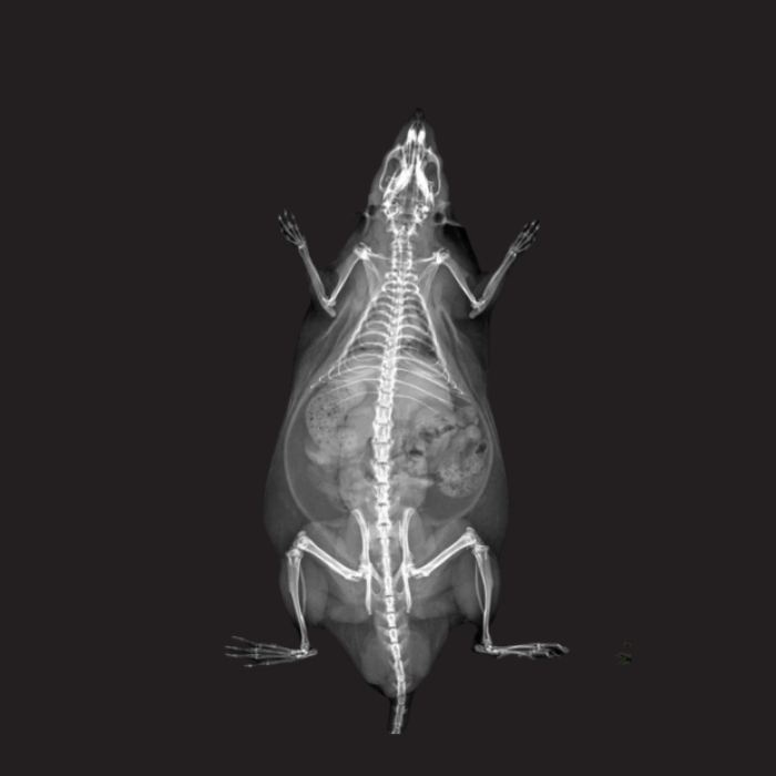
DXA Image Outputs
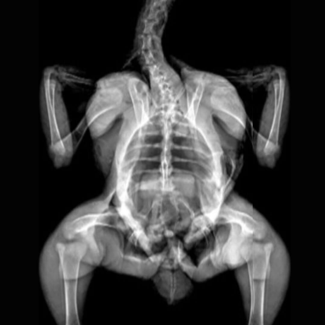
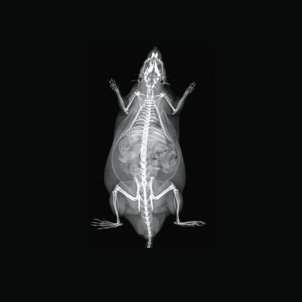
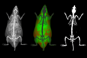
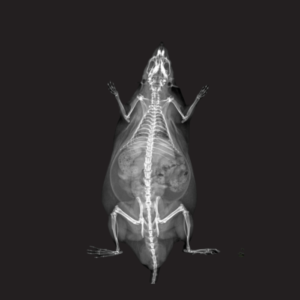
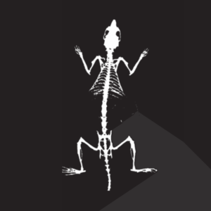
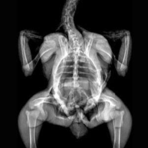
A single scan generates the X-ray attenuation image, bone mineral density map, and color map.
Applications in Preclinical Research
iNSiGHT DXA supports longitudinal assessment of bone and body composition across a range of preclinical disease models and study designs.
Metabolic Disorders
Metabolic disorders such as diabetes, metabolic syndrome, and obesity can alter body composition and musculoskeletal health. Small-animal models are commonly used to investigate these changes over time.
iNSiGHT DXA supports longitudinal assessment of fat mass, lean mass, bone mineral density, bone mineral content, and related body-composition endpoints, helping researchers characterize disease-associated changes or treatment response within the same study.
Measurements Relevant to Metabolic Research
- Fat mass and percentage
- Lean/non-bone mass and percentage
- Bone mineral content
- Total mass
- Longitudinal changes over time

Diabetic mouse model
In the cited study, streptozotocin-induced diabetic mice showed persistent decreases in fat mass, lower-limb lean mass, and bone mineral content measured by DXA.
Kishi et al., Exp Anim. 2023 · View study
Drug Safety & Toxicology
Drug safety and toxicology studies may include skeletal or body-composition endpoints when treatment-related changes in bone, fat, or lean tissue are relevant.
In those studies, iNSiGHT DXA can longitudinally quantify bone mineral density, bone mineral content, fat mass, and lean mass as part of a broader safety or treatment-response assessment.
Measurements Relevant to Drug Safety & Toxicology
- Bone mineral density
- Bone mineral content
- Fat mass
- Lean mass
- Longitudinal changes over time
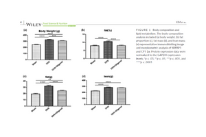
Ovariectomized rat model
In the cited study, iNSiGHT DXA was used in ovariectomized rats to measure body weight, bone mineral density, fat mass, fat proportion, and lean mass during oligonol treatment. The DXA measurements were used alongside molecular and histological assays to assess treatment-related changes.
Kim et al., Food Science & Nutrition. 2022 · View study
Musculoskeletal Diseases
Musculoskeletal research includes conditions and interventions affecting bone or muscle, including fracture and healing, bone or muscle regeneration, and muscle wasting.
iNSiGHT DXA can longitudinally quantify bone mineral density, bone mineral content, fat mass, and lean mass when those endpoints are relevant to disease progression, recovery, or treatment response.
Measurements Relevant to Musculoskeletal Research
- Bone mineral density
- Bone mineral content
- Fat mass
- Lean mass
- Longitudinal changes over time
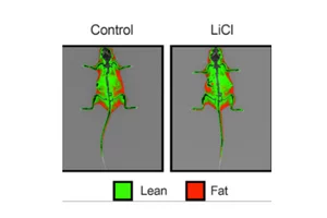
Low-dose lithium supplementation
In the cited study, OsteoSys iNSiGHT DXA was used to measure body composition in anesthetized mice during lithium treatment, while muscle and adipose mechanisms were evaluated with complementary assays.
Geromella et al., J Biol Chem. 2022 · View study
Metabolic Bone Disease & Arthritis
Metabolic bone diseases include conditions that affect bone mineralization or structure, including osteoporosis, rickets, osteomalacia, and osteogenesis imperfecta. Arthritis includes joint diseases such as osteoarthritis and rheumatoid arthritis.
iNSiGHT DXA can assess longitudinal changes in bone mineral density and bone mineral content across the whole animal or user-defined regions of interest, supporting metabolic bone studies and selected arthritis models where mineral endpoints are relevant.
Measurements Relevant to Bone & Joint Research
- Bone mineral density
- Bone mineral content
- Regional BMD and BMC within user-defined regions of interest
- Whole-body BMC and BMD
- Lumbar vertebral BMC and BMD
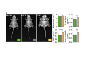
C3H mouse model
In the cited example, iNSiGHT DXA revealed higher BMC and BMD values in the whole body and lumbar vertebrae of C3H mice.
Kim MY et al., Exp Mol Med. 2021 · View study
Hypoxia
Hypoxic exposure in vivo can alter body composition and metabolic responses. Small-animal models are used to study these changes under defined hypoxic conditions.
iNSiGHT DXA can measure fat and lean body-composition endpoints across hypoxia protocols. Metabolic variables such as energy expenditure and substrate oxidation require complementary measurement methods.
Measurements Relevant to Hypoxia Research
- Fat mass
- Lean mass
- Body-composition changes across hypoxic exposure
- Longitudinal body-composition assessment

Hypoxic exposure in mice
In the cited study, DXA showed reduced body fat after 3 weeks of hypoxic exposure. Energy expenditure and carbohydrate and fat oxidation were measured separately using open-circuit calorimetry, not by DXA.
Park et al., BioMed Research International. 2020 · View study

Preclinical Imaging Workflow & System Features
iNSiGHT DXA combines streamlined animal handling, fast acquisition, and flexible imaging settings to support longitudinal preclinical studies.
Easy Data
Acquisition
Flexible Animal & Sample Range
25-Second
Scan Time
Low-Dose Longitudinal Imaging
Adjustable Field of View & Resolution
Shielded, Streamlined Workflow
Configuration & Add-ons
Configure iNSiGHT for Your Workflow
iNSiGHT DXA is available with optional add-ons to support anesthesia, higher-resolution imaging, and offline analysis.
Anesthesia Control
Anesthesia Control is available as an add-on for imaging workflows. Scintica also offers anesthesia systems for small-animal research.
Digital Radiography Mode
Digital Radiography Mode provides a pixel size of 31 µm at 4× magnification.
Offline Analysis Software
Available as an add-on for iNSiGHT DXA. For broader biomedical image analysis, Scintica also offers Imalytics Preclinical.
Need help building the right imaging workflow?
Scintica offers complementary small-animal anesthesia and biomedical image-analysis solutions alongside iNSiGHT DXA. Explore the related systems below or contact our team to discuss your study requirements.
Technical Specifications
Key imaging, animal-size, and system requirements for the iNSiGHT DXA platform
| Imaging & Acquisition | |
|---|---|
|
Main Use Case |
Preclinical in vivo DXA/DEXA for bone and body composition measurements |
|
Modality / Capabilities |
DXA/DEXA; bone mineral and body composition analysis |
|
Scan Method |
Cone Beam |
|
Pixel Size |
100 µm at 1.2X; 31 µm at 4X with Digital Radiography Mode |
| Animal & Scan Area | |
|---|---|
|
Animal Size |
Mice, rats, and animals of similar size (up to 500 g) |
|
Maximum Image Area |
16.5 × 25.5 cm |
| System Requirements | |
|---|---|
|
Operating System |
Windows 10 and 11, 64-bit |
|
Dimensions (W × D × H) |
66 × 61 × 113 cm |
|
Power |
110/240 VAC, 50/60 Hz, 200 VA |
|
Operating Temperature |
10–40 °C |
iNSiGHT DXA Imaging Gallery
Explore representative iNSiGHT DXA outputs from preclinical models, including X-ray attenuation images, body-composition color images, and bone mineral density maps across rat, marmoset, and chicken studies.


Continue Exploring iNSiGHT
Access product documentation and explore webinars, publications, application notes, and articles related to iNSiGHT DXA and preclinical bone and body-composition imaging.
Learn More About iNSiGHT DXA
Explore webinars, application-focused articles, and related research highlighting iNSiGHT DXA workflows and preclinical bone and body-composition studies.
(March 14, 2024) Webinar: Validation of DEXA for Longitudinal Quantification of Tumor Burden in A Murine Model of Pancreatic Ductal Adenocarcinoma
(March 12, 2026) Webinar: Preclinical Imaging of Multiple Myeloma Bone Disease by Dual-Energy X-ray Absorptiometry (DXA)
(March 14, 2024) Webinar: Validation of DEXA for Longitudinal Quantification of Tumor Burden in A Murine Model of Pancreatic Ductal Adenocarcinoma
Article: Wheat Seedling Extract Ameliorates Sarcopenia in Aged Mice by Regulating Protein Synthesis and Degradation with Anti-Inflammatory and Mitochondrial Biogenesis Effects
iNSiGHT DXA FAQ
Common questions about iNSiGHT DXA measurements, scan workflow, imaging outputs, and available system options.
What does iNSiGHT DXA measure?
iNSiGHT DXA provides quantitative measurements of bone mineral density (BMD), bone mineral content (BMC), bone area, tissue area, fat mass and percentage, lean mass and percentage, and total mass. Specific bone-length measurements can also be made from the 2D X-ray image. Analysis can be performed across the whole animal or within user-defined regions of interest (ROI).
What animal sizes are supported by iNSiGHT DXA?
The current iNSiGHT DXA page states an animal/sample range of 10 g to 500 g, with a maximum scan area of 16.5 × 25.5 cm. The Technical Specifications specifically describe use with mice, rats, and animals of similar size up to 500 g.
How long does an iNSiGHT DXA scan take?
A standard iNSiGHT DXA acquisition takes 25 seconds. In that scan, the system can generate the imaging outputs used for bone and body-composition analysis.
What images does iNSiGHT DXA generate?
A single scan generates an X-ray attenuation image, bone mineral density map, and color map. These outputs support quantitative bone and body-composition measurements for the whole animal or selected regions of interest.
What add-ons are available for iNSiGHT DXA?
The current iNSiGHT DXA configuration includes three listed add-ons:
- Anesthesia Control
- Digital Radiography Mode
- Offline Analysis Software
Ready to get started?
Ready to Discuss Your Preclinical DXA Workflow?
Have a question about whether iNSiGHT DXA is the right fit for your study? Our team can help you think through your imaging needs, study design, and system configuration.