

YOU MIGHT BE INTERESTED
InCiTe™ 3D X-Ray Microscope:
State-of-the-art benchtop micro-CT platform designed to deliver high-resolution, three-dimensional visualization of biological tissues, bone microarchitecture, and advanced materials.
Learn more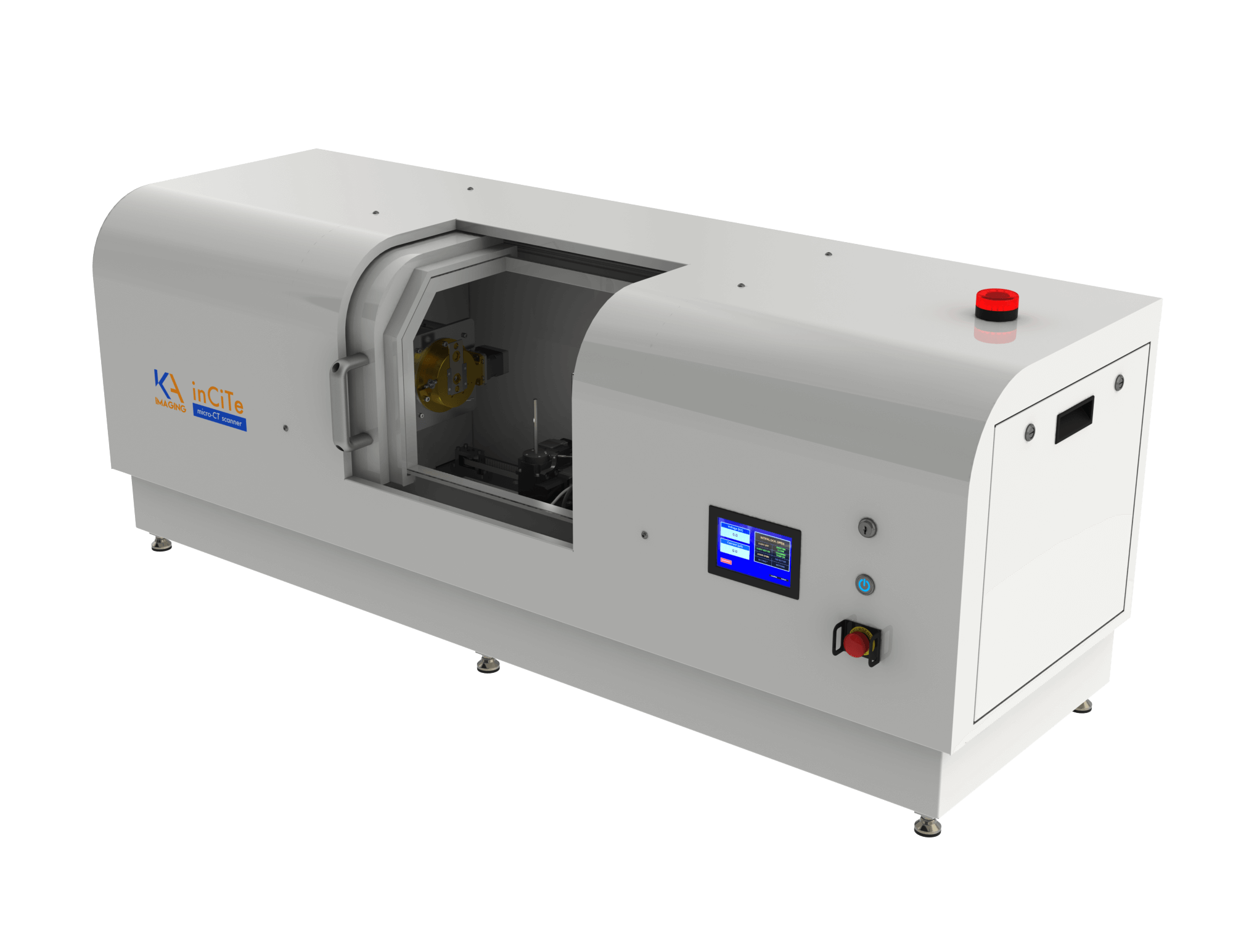
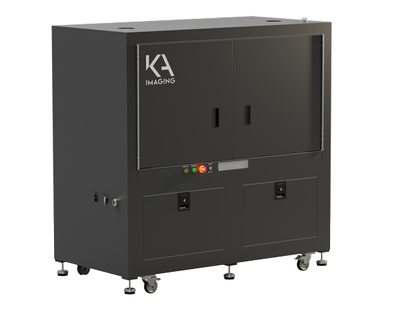
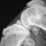
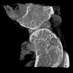
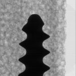
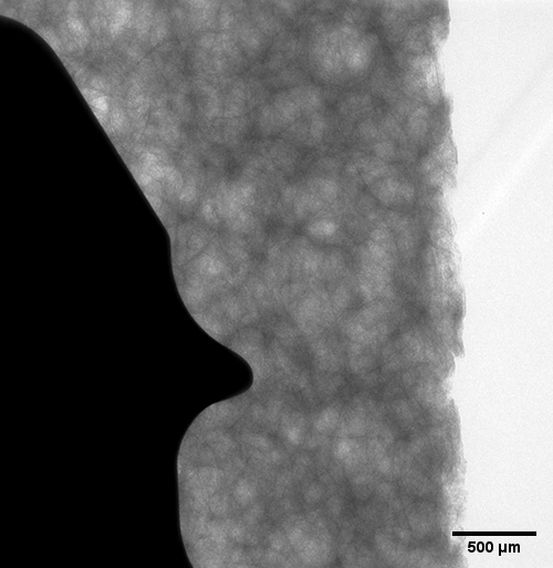
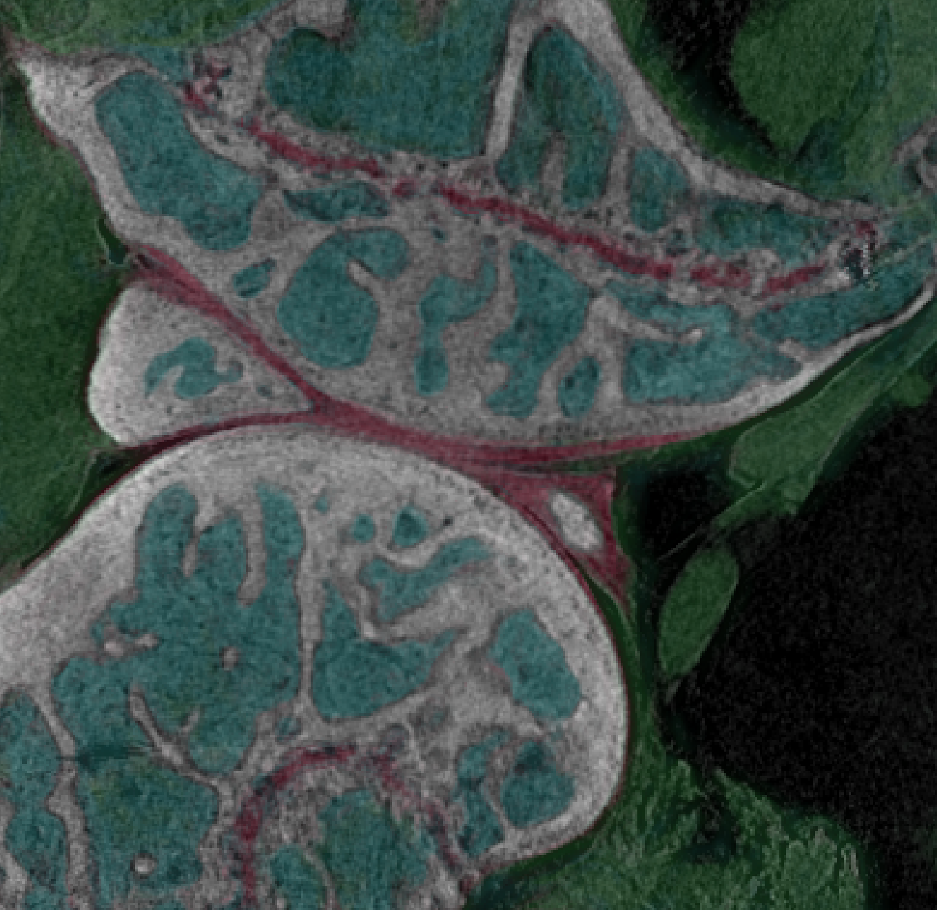
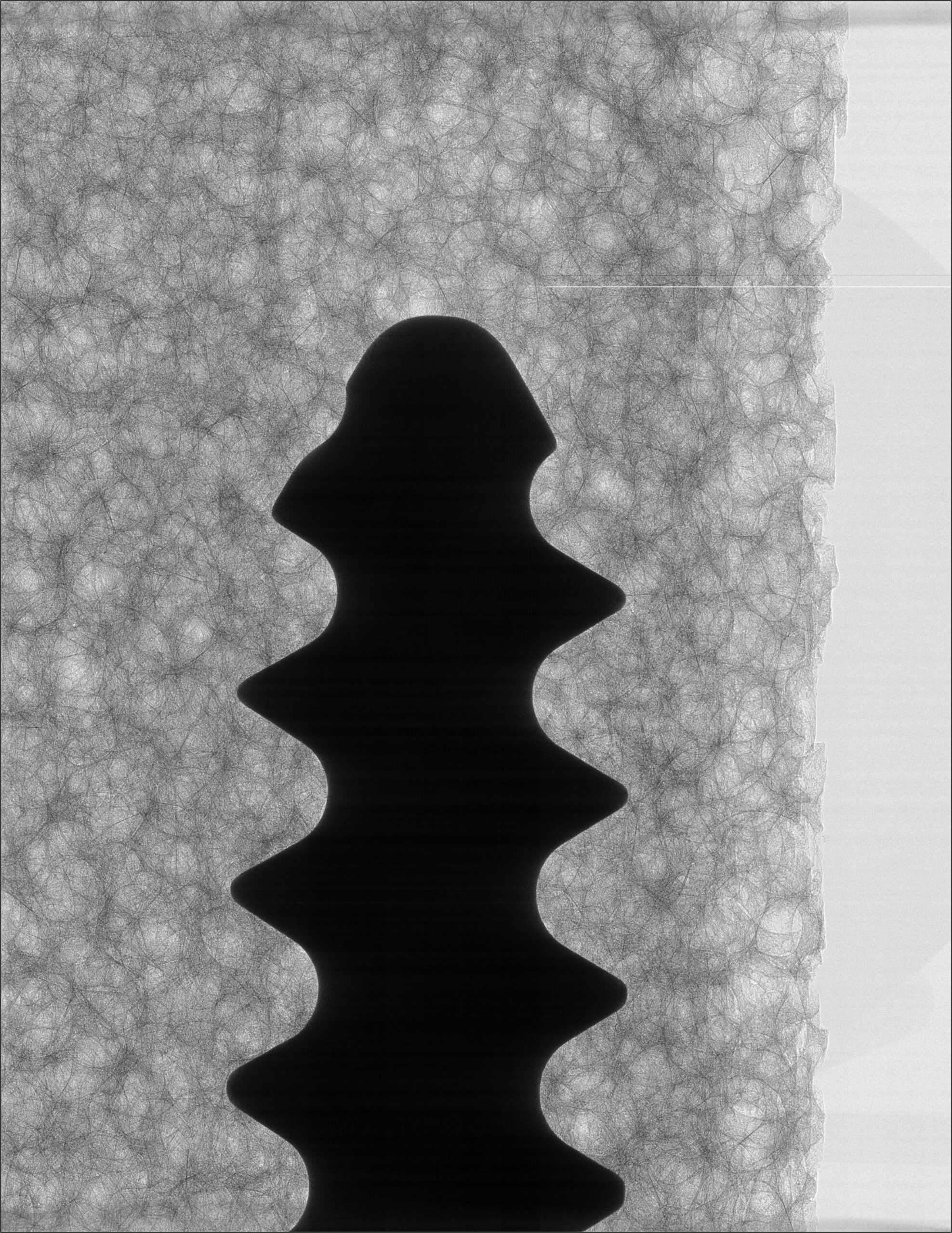
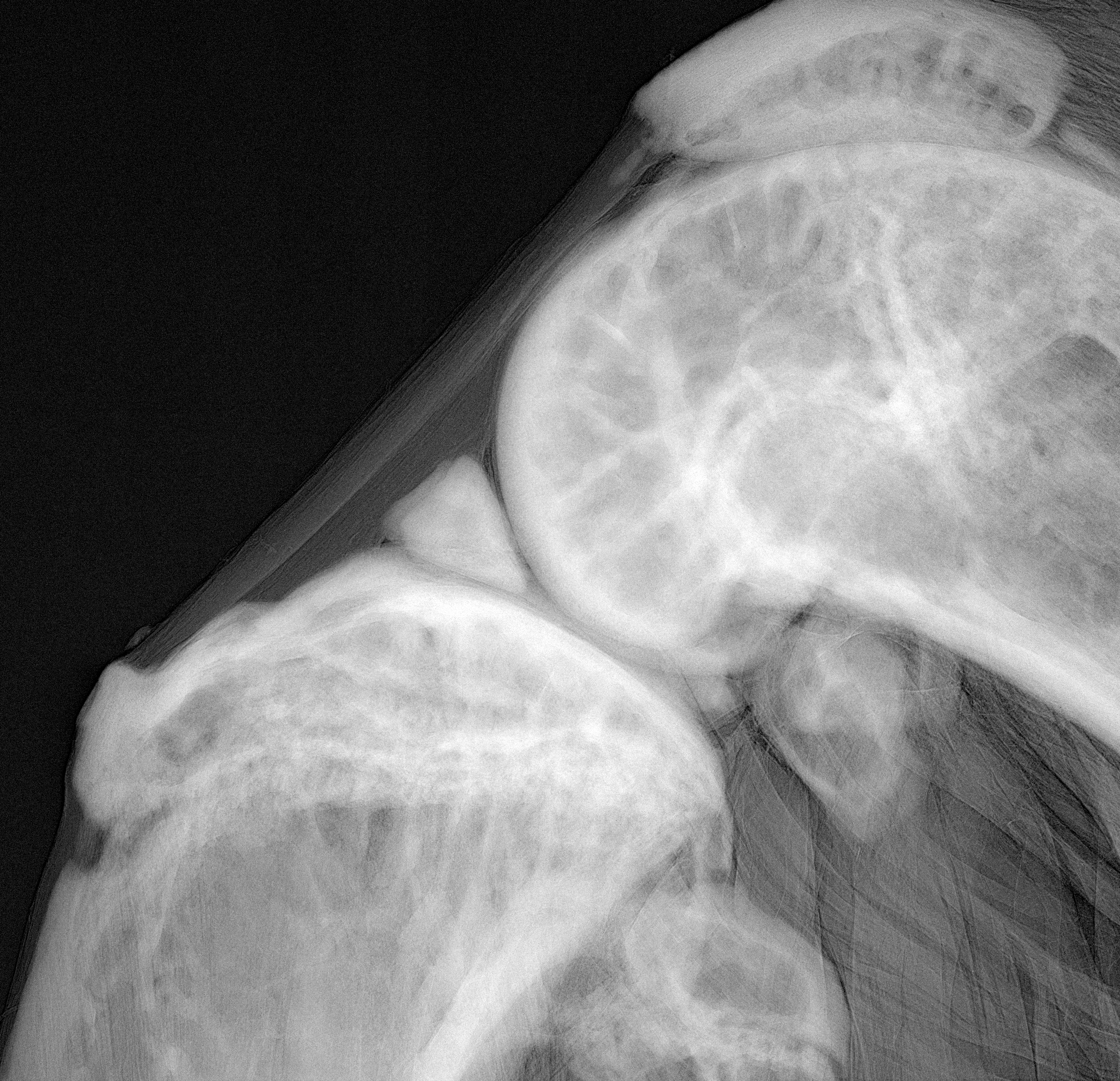
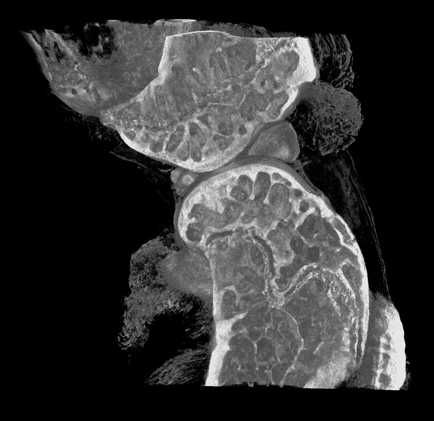
The inCiTe™ 2.0 3D X-ray Microscope is an advanced micro-CT system engineered for versatility. The system features an expanded internal cabinet to accommodate larger samples, supporting a variety of applications. Configured to perform phase contrast or spectral imaging, complex biological and orthopaedic structures can be investigated with confidence.
Re-configurable modular design and upgradable for maximum flexibility
Sub-micron pixel size at maximum magnification with the world-first BrillianSeTM a-Se/CMOS hybrid detector
810 mm (W) x 1780 mm (L) x 1800 mm (H)
1380 kg
100-240 VAC, 50-60 Hz
| BrillianSeTM Hybrid Detector | RevealTM Flat-Panel Detector | |
|---|---|---|
|
Pixel size at minimum magnification |
8 μm |
140 μm |
|
Field of view (FOV) |
30 mm (⌀) x 30 mm (H) |
300 mm (⌀) x 200 mm (H) |
|
FOV at maximum magnification |
3 mm (⌀) x 3 mm (H) |
10.7 mm (⌀) x 7.1 mm (H) |
|
Pixel size at maximum magnification |
0.8 μm (10x) |
5 μm (28x) |
*Specifications are preliminary and subject to change without notice.
YOU MIGHT BE INTERESTED
State-of-the-art benchtop micro-CT platform designed to deliver high-resolution, three-dimensional visualization of biological tissues, bone microarchitecture, and advanced materials.
Learn more