


Metabolic Disorders
Drug safety and toxicology
Musculoskeletal diseases
Metabolic Bone Disease & Arthritis
Hypoxia
Metabolic Disorders

Drug safety and toxicology
Musculoskeletal diseases
Metabolic Bone Disease & Arthritis
Hypoxia

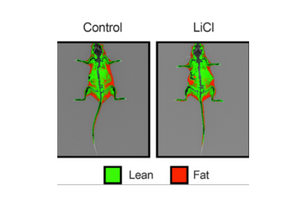
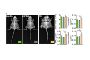
Research Beyond the PIXImus: Considerations for Replacing a Legacy Small Animal DXA System
Research Beyond the PIXImus: Considerations for Replacing a Legacy Small Animal DXA System
The Synergistic Power of Intermittent Fasting and Threonic Acid
Publication Highlight: The Synergistic Power of Intermittent Fasting and Threonic Acid in DIO

Article: Wheat Seedling Extract Ameliorates Sarcopenia in Aged Mice by Regulating Protein Synthesis and Degradation with Anti-Inflammatory and Mitochondrial Biogenesis Effects
Publication Highlight: Wheat Seedling Extract Ameliorates Sarcopenia in Aged Mice by Regulating Protein