Utilizing Ultrasound Imaging & Shear Wave Elastography to Evaluate Therapy Response of Tumors at an Early Stage
Ultrasound imaging combined with shear wave elastography (SWE) is an advanced technique increasingly used in clinical and pre-clinical research for assessing tumors. This method integrates the high-resolution imaging capabilities of traditional ultrasound with the biomechanical assessment provided by SWE, offering a comprehensive tool for non-invasive tumor evaluation.
Ultrasound imaging is widely utilized in both clinical and pre-clinical settings due to its ability to provide real-time, high-resolution images of soft tissues. It uses high-frequency sound waves that penetrate the body, reflect off tissues, and return to the transducer, creating detailed images based on the differing acoustic properties of tissues. In tumor research, ultrasound imaging allows for the visualization of tumor size, shape, location, and vascularity, enabling researchers to monitor tumor growth and progression over time. Shear wave elastography is a functional imaging technique that measures the stiffness or elasticity of tissues. It works by generating mechanical shear waves within the tissue using acoustic radiation force. These waves propagate through the tissue, and their speed is directly related to the tissue’s stiffness. The ultrasound transducer detects these waves and calculates the elasticity modulus, producing a quantitative map (elastogram) of tissue stiffness.
In the context of tumors, tissue stiffness is a crucial biomarker. Malignant tumors are often stiffer than benign tumors or normal tissues due to increased cell density, fibrosis, and changes in the extracellular matrix. Therefore, SWE can provide valuable insights into the biomechanical properties of tumors, which are often indicative of tumor type, aggressiveness, and stage.
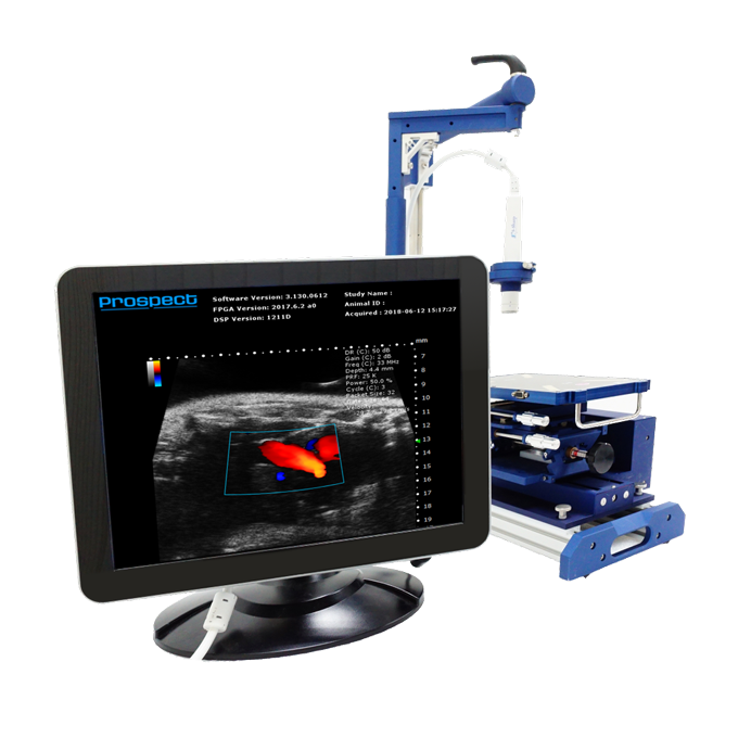
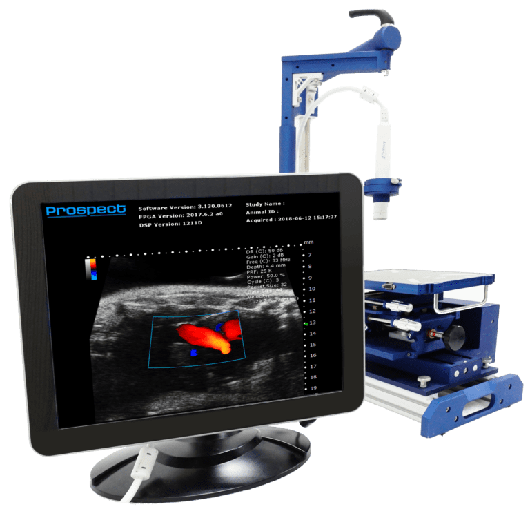
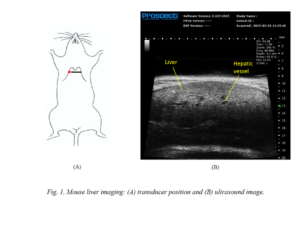
*All data shown was acquired from Dr. Jason Holland’s lab using the Prospect T1 High-frequency Ultrasound System

Applications in Preclinical Research
In preclinical research, particularly in oncology, the combination of ultrasound imaging with SWE offers several advantages:
- Tumor Detection and Characterization:
- Structural Imaging: Ultrasound provides detailed images of the tumor’s structure, allowing researchers to detect and monitor tumors in animal models. It helps in assessing parameters like tumor size, shape, and growth dynamics.
- Elasticity Mapping: SWE adds a layer of functional information by mapping tissue stiffness. This is particularly useful for differentiating between malignant and benign tumors, as well as identifying tumor margins, which can be challenging with conventional imaging alone.
- Monitoring Tumor Progression:
- Longitudinal Studies: The non-invasive nature of ultrasound and SWE allows for repeated measurements in the same animal over time, making it ideal for longitudinal studies. Researchers can track changes in tumor stiffness as the tumor progresses or regresses, providing insights into the natural history of the tumor.
- Tumor Growth and Invasion: Changes in tissue stiffness can be correlated with tumor growth and invasiveness. As tumors become more aggressive, they often become stiffer, and this can be monitored using SWE.
- Evaluating Treatment Response:
- Therapeutic Efficacy: SWE can be used to assess the effectiveness of anti-cancer treatments by monitoring changes in tumor stiffness. For example, effective therapies may reduce tumor stiffness, indicating a reduction in tumor density or fibrosis.
- Early Detection of Treatment Response: Changes in elasticity may occur before changes in tumor size are detectable by conventional ultrasound, providing an early indicator of treatment efficacy.
- Studying Tumor Microenvironment:
- Biomechanical Properties: The tumor microenvironment, including the extracellular matrix, plays a crucial role in cancer progression. SWE can help in studying the biomechanical properties of the tumor microenvironment, offering insights into how these properties influence tumor behavior and response to therapy.
- Modeling Tumor Behavior: By combining structural and stiffness data, researchers can develop more accurate models of tumor behavior, which can be used to predict outcomes or tailor treatment strategies in pre-clinical models.


Advantages of Using Ultrasound with SWE in Preclinical Research
- Non-Invasiveness: Both ultrasound imaging and SWE are non-invasive, reducing the need for biopsies and minimizing harm to animal subjects.
- Real-Time Monitoring: These techniques provide real-time data, allowing for immediate assessment and decision-making in experimental studies.
- Quantitative Data: SWE provides quantitative measurements of tissue stiffness, which can be objectively analyzed and compared across studies.
- Cost-Effectiveness: Ultrasound and SWE are relatively cost-effective compared to other imaging modalities like MRI or CT, making them accessible for routine use in pre-clinical research.

Conclusion
Ultrasound imaging combined with shear wave elastography is a versatile and powerful tool in preclinical tumor research. It enables detailed assessment of tumor structure and biomechanics, offering valuable insights into tumor characterization, progression, and response to therapy. This non-invasive approach facilitates longitudinal studies and reduces the need for invasive procedures, making it an essential technique for advancing cancer research and developing new therapeutic strategies.
References
- Cosgrove, D. O., et al. (2013). Shear wave elastography for breast masses is highly reproducible. European Radiology, 23(12), 3194-3202.
- Barr, R. G. (2015). Shear wave liver elastography. Abdominal Imaging, 40(4), 692-707.
- Gennisson, J. L., Deffieux, T., Fink, M., & Tanter, M. (2013). Ultrasound elastography: principles and techniques. Diagnostic and Interventional Imaging, 94(5), 487-495.
- Cantisani, V., et al. (2015). Ultrasound elastography in the evaluation of liver, prostate, and thyroid diseases. European Journal of Radiology, 84(4), 611-617.
- Liu, B., Zheng, Y., Huang, G., Lin, M., Shan, Q., & Lu, Z. (2017). Shear wave elastography in the diagnosis of thyroid nodules: a systematic review and meta-analysis. Scientific Reports, 7(1), 4669.
- Bracco, C., & Pretolesi, F. (2017). Ultrasound Elastography for the characterization of focal liver lesions. Insights into Imaging, 8(5), 1-11.

Watch Our Webinars!
Articles & Publications
Recent Posts
Prospect T2