


Functional Connectivity
Task-Based Functional Imaging
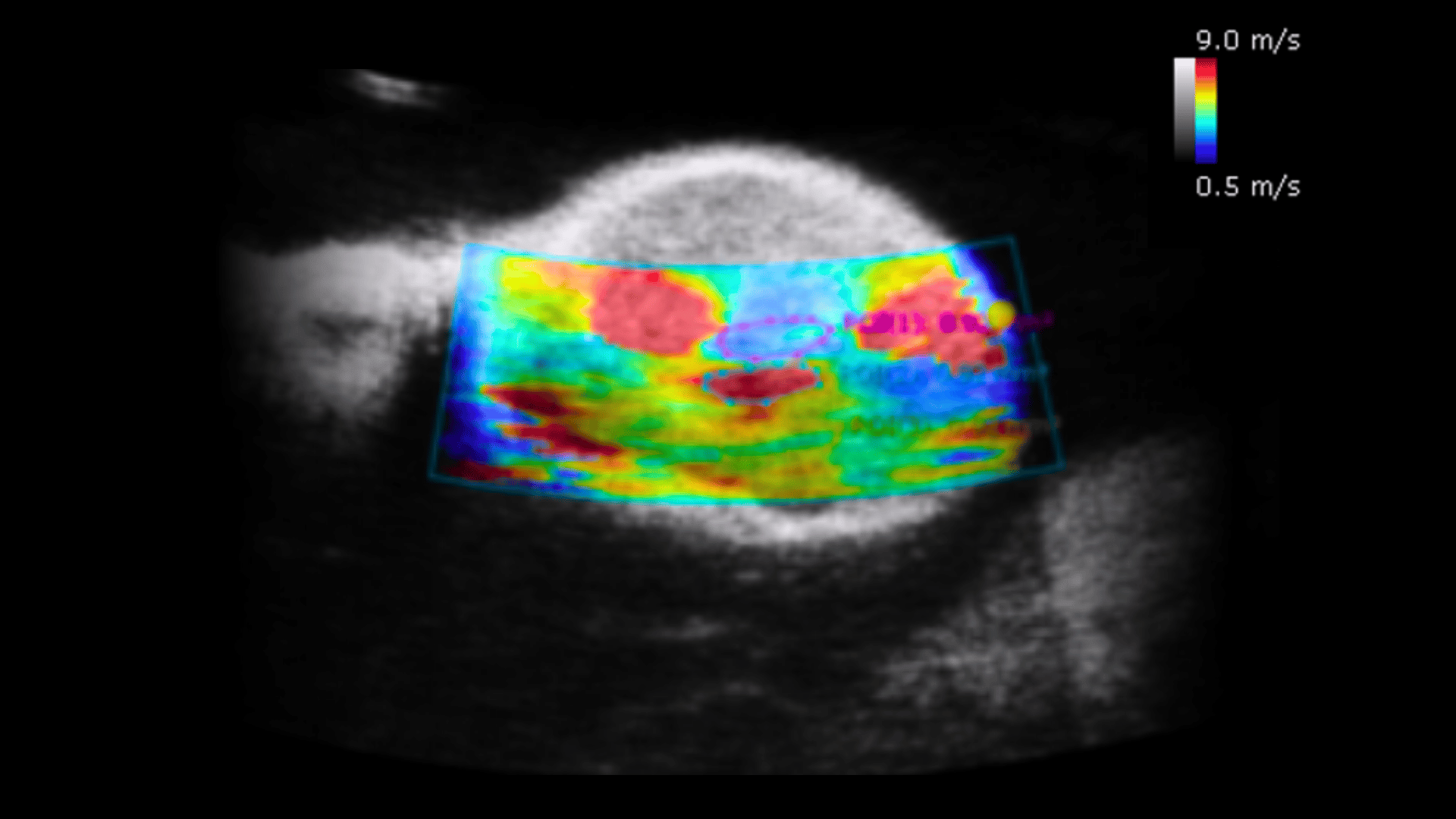
Stroke & Vascular imaging
Neuropharmacology
Super-Resolution Ultrasound Localization Microscopy (ULM)
Functional Connectivity

Task-Based Functional Imaging

Stroke & Vascular imaging

Neuropharmacology

Super-Resolution Ultrasound Localization Microscopy (ULM)


Clinical vs. Preclinical Ultrasound: Why Frequency Matters
Clinical vs. Preclinical Ultrasound: Why Frequency Matters View Paper Here Our Ultrasound SystemProspect

Scintica Morning Coffee Chat Episode 1: DXA & Ultrasound
https://youtu.be/KfbEW_b5Gpg?si=2xXwoxw-DuGtjq77 Join us on our first episode of the Scintica Morning Coffee Chat. This

Science Corner: Utilizing Ultrasound Imaging & Shear Wave Elastography to Evaluate Therapy Response of Tumors at an Early Stage
Utilizing Ultrasound Imaging & Shear Wave Elastography to Evaluate Therapy Response of Tumors