Preclinical Contrast Agents vs Clinical Agents in Small Animal Imaging
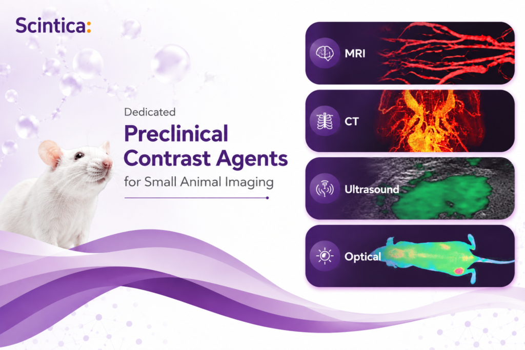
Preclinical contrast agents optimized for small animal imaging can help researchers better align signal behavior, circulation time, dosing, and modality-specific endpoints across MRI, CT, ultrasound, and optical imaging. Compared with clinical agents designed for human diagnostic imaging, dedicated preclinical agents are developed around rodent physiology, small injection volumes, and the practical needs of longitudinal imaging studies.
Why Dedicated Preclinical Contrast Agents Matter in Small Animal Imaging
Contrast agents can strongly influence what researchers are able to see, quantify, and interpret in preclinical imaging. They help define vascular structures, improve tissue contrast, support functional imaging, and make subtle biological changes easier to detect. However, not every contrast agent is designed for the same biological setting.
Clinical contrast agents are developed for human diagnostic imaging. Small animal studies work under different constraints: smaller blood volume, faster metabolism, narrow dosing margins, high resolution imaging requirements, and often longitudinal imaging for the same cohort. For preclinical research, the question is not simply whether a clinical agent can produce contrast. The more useful question is whether that agent matches the animal model, imaging modality, and biological endpoint.
Dedicated preclinical contrast agents are designed around small animal imaging workflows. They can help researchers align pharmacokinetics, signal behavior, dosing, circulation time, and documentation with the needs of rodent imaging studies.
Key Takeaways
- Clinical contrast agents are optimized for human imaging and may not translate predictably to small animal studies.
- Preclinical agents are developed with rodent physiology, small injection volumes, and small imaging targets in mind.
- Agent selection can affect signal intensity, circulation time, biodistribution, tolerability, and longitudinal study design.
- The right contrast strategy should be matched to the modality, endpoint, animal model, and imaging window.
Why Clinical Agents May Not Be the Best Fit
Using a familiar clinical agent in a preclinical study can seem convenient, but small animal imaging is not just clinical imaging at a smaller scale. A rodent study may require a different concentration, injection volume, imaging window, and clearance profile than a clinical workflow.
If an agent clears too quickly, the imaging window may be too short. If the signal is not strong enough for a small target, subtle vascular, tumor, organ, or biodistribution findings may be harder to interpret.
These issues are especially important in longitudinal studies, where researchers follow disease progression, treatment response, or functional change. In these workflows, consistency matters as much as contrast.
How Preclinical Contrast Agents Support Study Design
| Study need | Why it matters in small animal imaging |
|---|---|
| Rodent matched pharmacokinetics | Supports more appropriate circulation, biodistribution, and clearance behavior for mice and rats. |
| High signal in small volumes | Helps improve visualization of small tumors, microvasculature, organ compartments, or functional targets. |
| Functional imaging windows | Blood pool or vascular agents can support perfusion, cardiac, and dynamic imaging questions. |
| Longitudinal compatibility | Relevant when repeated dosing, survival imaging, tolerability, and animal welfare are part of the study plan. |
| Preclinical documentation | Datasheets and protocols can reduce uncertainty during study setup and improve repeatability. |
Preclinical vs Clinical Contrast Agents
| Feature | Dedicated preclinical agents | Clinical contrast agents |
|---|---|---|
| Designed around rodent physiology | Yes | Not typically |
| Optimized for small imaging targets | Yes | Not typically |
| Targeted or molecular options | Often available | Limited for preclinical endpoints |
| Preclinical protocols and documentation | Yes | Often limited |
| Fit for repeated small animal imaging | Can be selected for this need | Requires careful validation |
Need help matching an agent to your imaging workflow?
Explore Viscover preclinical contrast agents or contact Scintica for support selecting an agent based on modality, animal model, imaging window, and biological endpoint.
Match the Agent to the Modality
A strong contrast agent strategy begins with the imaging endpoint. MRI, CT, ultrasound, and optical imaging each provide different insights and require different contrast behavior. A study focused on vascular ultrasound has different needs than a CT study for organ enhancement, an MRI study of soft tissue contrast, or an optical imaging study focused on biodistribution.
This is why contrast agents should be selected as part of the experimental design, alongside the animal model, imaging modality, timing, anesthesia workflow, and analysis plan.
| Modality | Typical contrast agent consideration |
|---|---|
| MRI | Soft tissue contrast, biodistribution, vascular or organ-specific enhancement. |
| CT | High-density or organ-targeted contrast for anatomical and structural visualization. |
| Ultrasound | Microbubble-based agents for perfusion, vascular, targeted, or dynamic imaging. |
| Optical Imaging | Fluorescent or near-infrared agents for biodistribution and molecular imaging workflows. |
Practical Workflow for Choosing an Agent
Define the biological question
Anatomy, perfusion, vascular change, tumor targeting, inflammation, biodistribution, or treatment response.
Select the modality and endpoint
Match the imaging method to the analysis endpoint and the type of data the study needs to generate.
Identify the imaging window
Consider immediate, vascular residence, delayed, or longitudinal follow up requirements.
Confirm animal model constraints
Species, body weight, route of administration, survival requirements, and dosing schedule.
Review protocol documentation
Review available datasheets and protocols before scaling to a full study.
Product Highlight: Viscover Preclinical Contrast Imaging Agents
Preclinical Contrast Imaging Agents
Modality-specific agents for small animal imaging workflows
Viscover provides a portfolio of preclinical contrast imaging agents for small animal research across MRI, CT, ultrasound, and optical imaging.
Designed for mice and rats, these agents simplify selection by matching the imaging method, animal model, and biological endpoint.


Related Resources
Frequently Asked Questions
Are clinical contrast agents unsuitable for every preclinical study?
Not necessarily. Clinical agents can sometimes be used, but they should not be assumed to be optimal without validation. The key is whether the agent fits the animal model, modality, imaging window, and endpoint.
What is the main advantage of a preclinical contrast agent?
The main advantage is better alignment with small animal imaging workflows, including dosing volume, circulation behavior, signal performance, documentation, and repeated imaging needs.
Which modalities can use dedicated preclinical agents?
Dedicated preclinical contrast agents are available for MRI, CT, ultrasound, and optical imaging. The best choice depends on the research question and the data the study needs to generate.
Final Thoughts
In small animal imaging, the contrast agent can affect the imaging window, signal quality, reproducibility, and biological interpretation of a study. Clinical contrast agents are built for human diagnostic imaging, while preclinical studies require closer alignment with rodent physiology and small scale imaging endpoints.
By choosing a dedicated preclinical contrast agent early in the study design process, researchers can build workflows that better match the modality, animal model, and scientific question.
Browse Preclinical Imaging Agents
Explore Scintica’s online collection of preclinical imaging agents for MRI, CT, ultrasound, and optical imaging workflows.
Browse Imaging Agents