A Century of Fighting Back Against Cancer
Explore the preclinical discoveries that have shaped how cancer is studied, modelled, and treated. This timeline highlights the scientific progress behind modern oncology research, from early tumour biology to emerging drug discovery workflows.


Preclinical Milestones
The Discoveries That Defined Cancer Science
Each breakthrough below began in a laboratory, with a hypothesis, a model, and the relentless pursuit of understanding.
Discovery of X-Rays
Wilhelm Röntgen discovers X-rays, launching radiation as both a diagnostic and therapeutic tool. Within years, researchers begin applying it to shrink tumours in preclinical models.
View ReferencePeyton Rous & the Tumour Virus
Rous demonstrates that a virus (RSV) can cause cancer in chickens, the first evidence that infectious agents drive malignant transformation. A Nobel Prize follows 55 years later.
View ReferencePapanicolaou Develops the Pap Test
George Papanicolaou discovers that cervical cancer can be detected by examining cells under a microscope, laying the groundwork for one of the most effective cancer-screening tools ever created.
View ReferenceWatson & Crick Describe DNA's Double Helix
The structure of DNA is unveiled, providing the molecular blueprint for all cancer biology. Understanding how genetic information is stored and mutated becomes the bedrock of oncology for generations.
View ReferencePhiladelphia Chromosome Identified
Nowell and Hungerford discover the first chromosomal abnormality linked to cancer in CML, the first proof that cancer has a definable genetic basis, opening the door to targeted therapy.
View ReferenceKnudson's Two-Hit Hypothesis
Alfred Knudson proposes that cancer requires two separate genetic mutations to develop, a foundational preclinical framework explaining how tumour-suppressor genes are inactivated and hereditary syndromes arise.
View ReferenceFirst Cellular Oncogene (src) Cloned
Bishop and Varmus show that oncogenes originate from normal cellular genes mutated by viruses or spontaneous changes. This Nobel-winning preclinical work reveals cancer is fundamentally a disease of the genome.
View Referencep53 Tumour Suppressor Discovered
The p53 gene, the most commonly mutated gene across all human cancers, is identified. Its protein controls cell proliferation and suppresses tumour growth, reshaping our understanding of cancer biology entirely.
View ReferenceHER2 Oncogene Characterised
HER2/neu is identified as a growth-factor receptor driving aggressive breast and ovarian cancers. Decades of preclinical work eventually leads to trastuzumab (Herceptin), transforming care for HER2-positive patients.
View ReferenceBRCA1 Gene Linked to Hereditary Breast Cancer
Researchers map the first locus for hereditary breast cancer, leading to BRCA1 cloning in 1994. This milestone enables genetic testing and prophylactic strategies that save thousands of lives annually.
View ReferenceAnti-Angiogenesis Therapy Validated
Folkman's hypothesis, that tumours require new blood vessel growth to survive, is vindicated when anti-VEGF antibodies dramatically suppress tumour growth in animal models, paving the way for bevacizumab.
View ReferenceHuman Genome Project Completed
The full mapping of human DNA unlocks the ability to identify the specific mutations powering cancer. This achievement catalyses every subsequent era of precision oncology and biomarker-driven drug discovery.
View ReferenceCAR-T Cell Technology Developed
Sadelain, Brentjens and Rivière begin engineering T cells with chimeric antigen receptors (CARs), creating genetically reprogrammed immune soldiers capable of seeking and destroying cancer cells with remarkable precision.
View ReferencePD-1/PD-L1 Checkpoint Pathway Elucidated
Preclinical work reveals how tumours exploit the PD-1 checkpoint to evade immune destruction, underpinning Nobel Prize–winning checkpoint immunotherapy, one of the most transformative shifts in cancer treatment history.
View ReferenceCRISPR-Cas9 Adapted for Genome Editing
Doudna and Charpentier adapt CRISPR-Cas9 into a precise gene-editing tool. In cancer research it rapidly enables identification of driver genes, engineering of tumour models, and next-generation cell therapy development.
View ReferencePatient-Derived Tumour Organoids Established
Researchers develop 3D self-organising tumour "mini-organs" grown from patient tissue. These organoids accurately replicate tumour architecture and heterogeneity, dramatically improving preclinical drug testing accuracy.
View ReferenceTargeted Protein Degradation (TPD) Emerges
PROTAC molecules that selectively degrade previously "undruggable" cancer proteins begin showing dramatic preclinical efficacy, opening a new therapeutic modality for targets long considered inaccessible.
View ReferenceFDA Modernization Act 2.0 Passed
New legislation allows drug approvals without mandatory animal testing, accelerating uptake of organoids, organs-on-chips, and AI models as primary preclinical evidence, a regulatory revolution for cancer drug development.
View ReferenceAI Transforms Preclinical Drug Discovery
Deep learning models predict immunotherapy responses, identify novel targets, and accelerate drug screening. AI-assisted imaging is now being validated across lung, breast, brain, and colorectal cancer preclinical pipelines.
View ReferenceTPD Enters Paediatric Brain Tumour Trials
A programme combining targeted protein degradation with personalised immunotherapy advances into clinical trials for paediatric brain and solid tumours, one of the first clinical examples of TPD in childhood cancers.
View ReferenceCancer research is not a sprint. It is a century-long relay each scientist passing the baton to the next
Cancer Awareness Month -2026
Every Breakthrough
Starts in a Lab
As we come to the end of Cancer Awareness Month, behind every patient outcome is a preclinical discovery. We want to honor the scientist who ask the questions no one else will.
Equipment Supporting
Cancer Research
The Prospect T2 is an innovative high-frequency ultrasound system designed specifically for in vivo preclinical imaging in small animals such as mice and rats. This compact and cost-effective tablet-based system provides high-resolution images (up to 30 µm) and advanced capabilities to monitor changes in hemodynamics and observe anatomical structures in real-time.
Allogeneic Expanded Human Peripheral NK Cells Control Prostate Cancer Growth in a Preclinical Mouse Model of Castration-Resistant Prostate Cancer


The M-Series™ systems are cryogen/cooling-free, self-shielded, high-performance MRI systems based on permanent magnet technology. They allow preclinical researchers to utilize the gold standard in soft tissue imaging without the cost, complexity, or technical burden of superconducting systems. Models support mice, rats, and non-human primate brains. With no special infrastructure requirements and minimal operating costs, these 1 Tesla systems are optimized for anatomical, functional, and molecular imaging in cancer, cardiac, neuroscience, and multimodal applications. The optional SimPET insert enables simultaneous PET/MRI acquisition.
Palbociclib Releases the Latent Differentiation Capacity of Neuroblastoma Cells


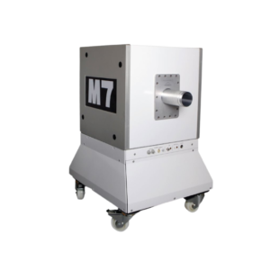
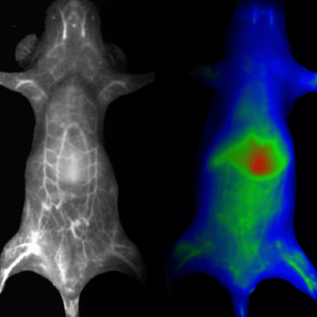
Newton is the first deeply cooled CCD imager dedicated to both fluorescence and bioluminescence multispectral imaging for a wide range of in vivo applications. Ideal for preclinical research, it provides non-ionizing and non-invasive visualization of biological processes in real time in the visible, near and short-wave infrared spectrum (VIS/NIR/NIR-II). Whether your focus is oncology, immunology, infectious disease, neurology, or biodistribution studies, our all-in-one system accommodates them all.
A Targeted Lipid Nanoparticle Platform for T Cell-Specific Non-Viral DNA Delivery & In Vivo CAR-T Generation


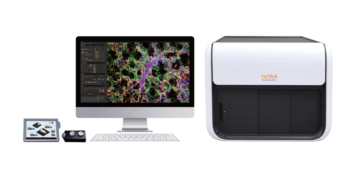
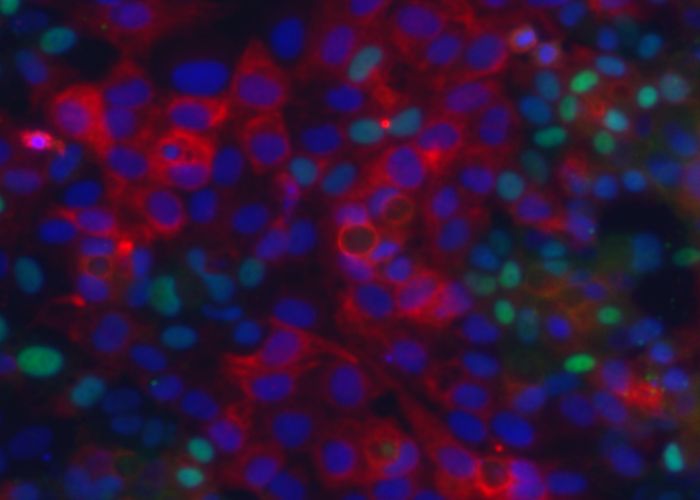
The IVM is an all-in-one two-photon and/or confocal microscopy system designed and optimized for longitudinal imaging of live animal models in vivo. Built around ease-of-use and augmented throughput, it is a next-generation core technology for biologists and translational scientists to elucidate the underlying mechanisms of biological phenomena at tissue and cellular level. Confocal IVM systems enable optical sectioning of in vivo tissue via rejection of out-of-focus fluorescence, resulting in images with high contrast and quality.
Inhalable siRNA Nanoparticles for Enhanced Tumor-Targeting Treatment of KRAS-Mutant Non-Small-Cell Lung Cancer


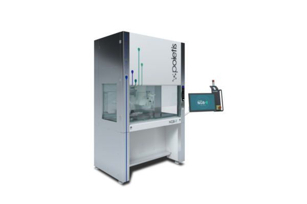
The NGB-R is a multimodal, 3D bioprinting platform designed and developed to print live tissues and organs. Combining laser-assisted, micro-valve, and extrusion bioprinting, the NGB-R enables true versatility of bioprinting (from cells to spheroids) and offers the possibility of using a large number of biomaterials and hydrogels.
Bioprinting Beyond the Limitations of Traditional Cancer Models


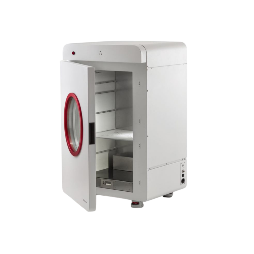
The InvivO2 workstation is packed with innovative features that allow you to study even the most complex cell interactions under perfect physiological oxygen conditions. Whether replicating the environment of blood vessels or lung tissue, the InvivO2 is the best tool for the job. Easy to use and adaptable for cell culture applications requiring carefully controlled oxygen regulation, it offers accurate and stable user-defined environmental controls with direct access to the inner chamber.
Nobel Prize for Physiology and Medicine 2019 Awarded for Discovery in Oxygen Related Mechanisms of Disease


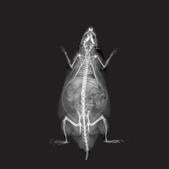
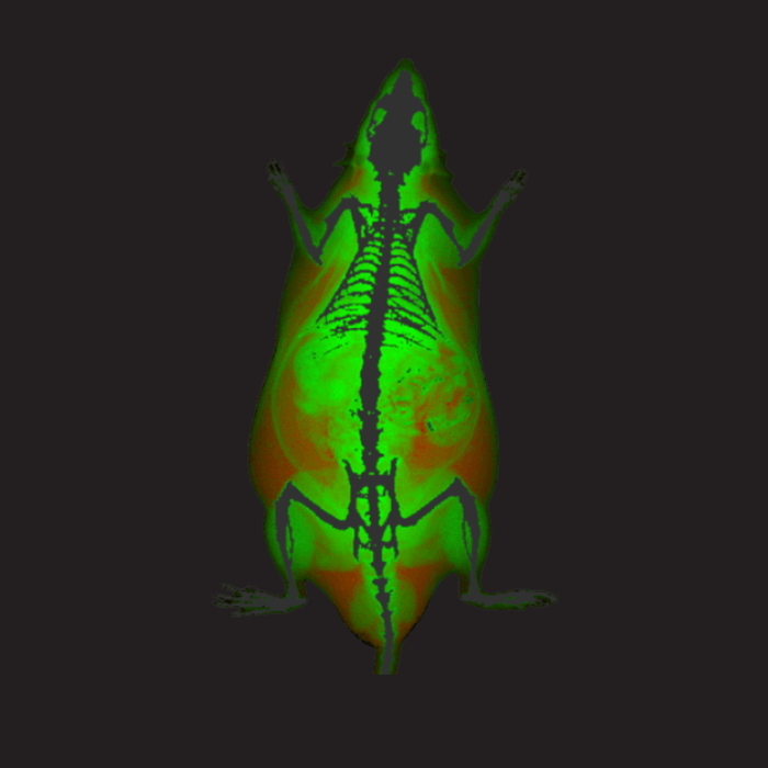
The iNSiGHT DXA system is a state-of-the-art in vivo Dual Energy X-Ray Absorptiometry (DXA/DEXA) technology designed for preclinical research. It provides a comprehensive solution for evaluating a wide range of metabolic disorders, such as osteoporosis, arthritis, diabetes, obesity, and musculoskeletal pathologies, including bone regeneration and muscle wasting diseases. Equipped with a fully shielded X-ray cabinet, it is suitable for small animal DXA applications.
Validation of DEXA for Longitudinal Quantification of Tumor Burden in A Murine Model of Pancreatic Ductal Adenocarcinoma

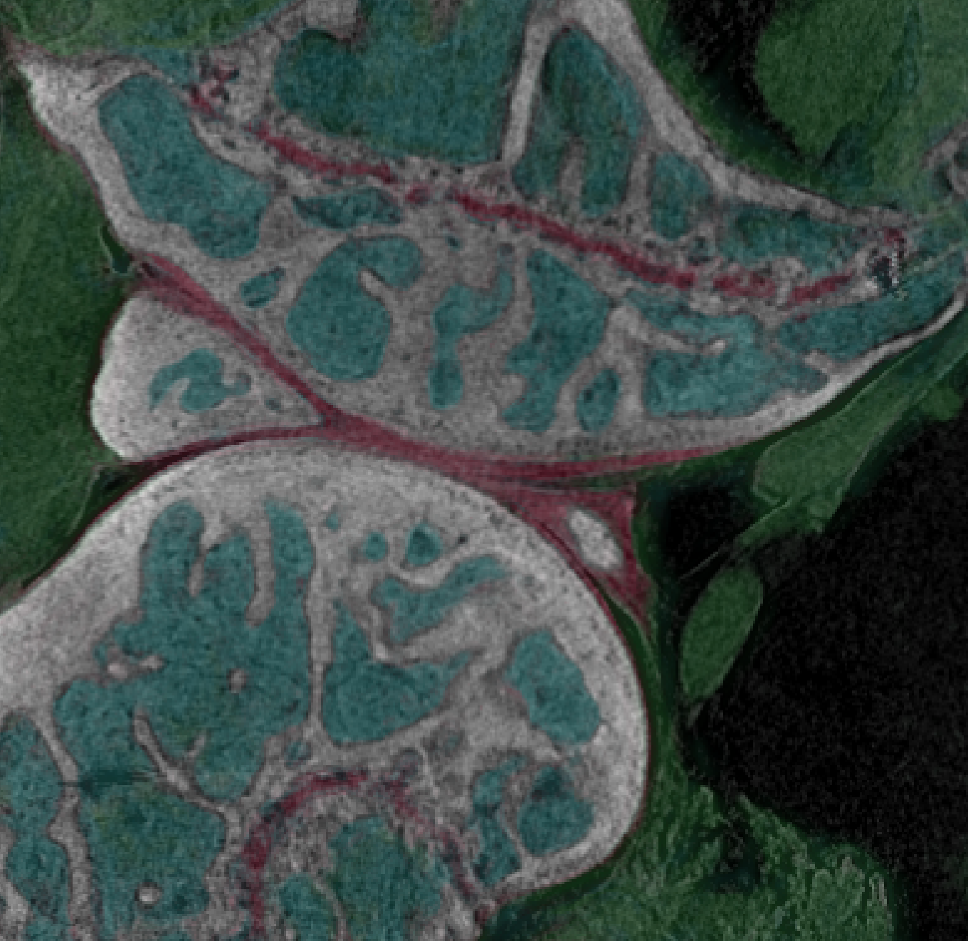
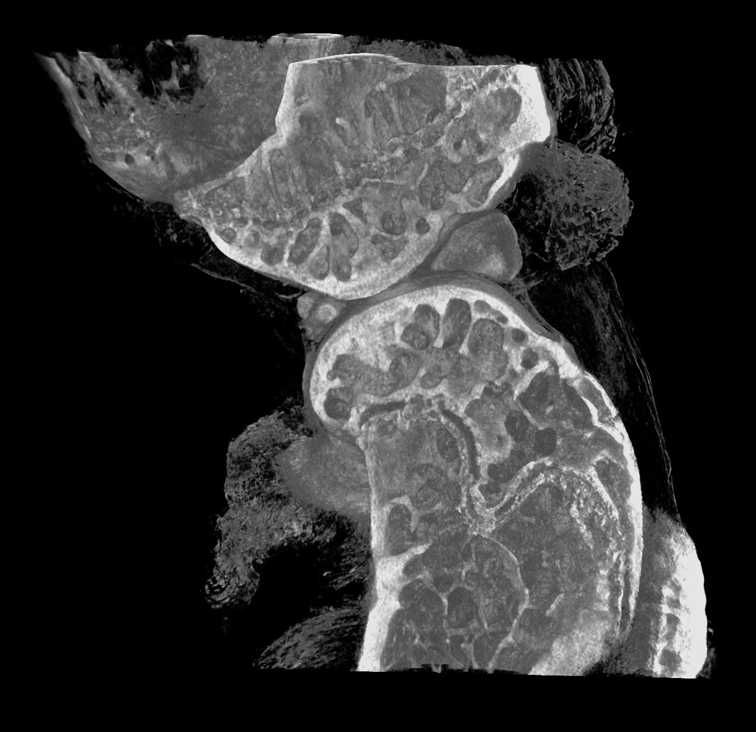
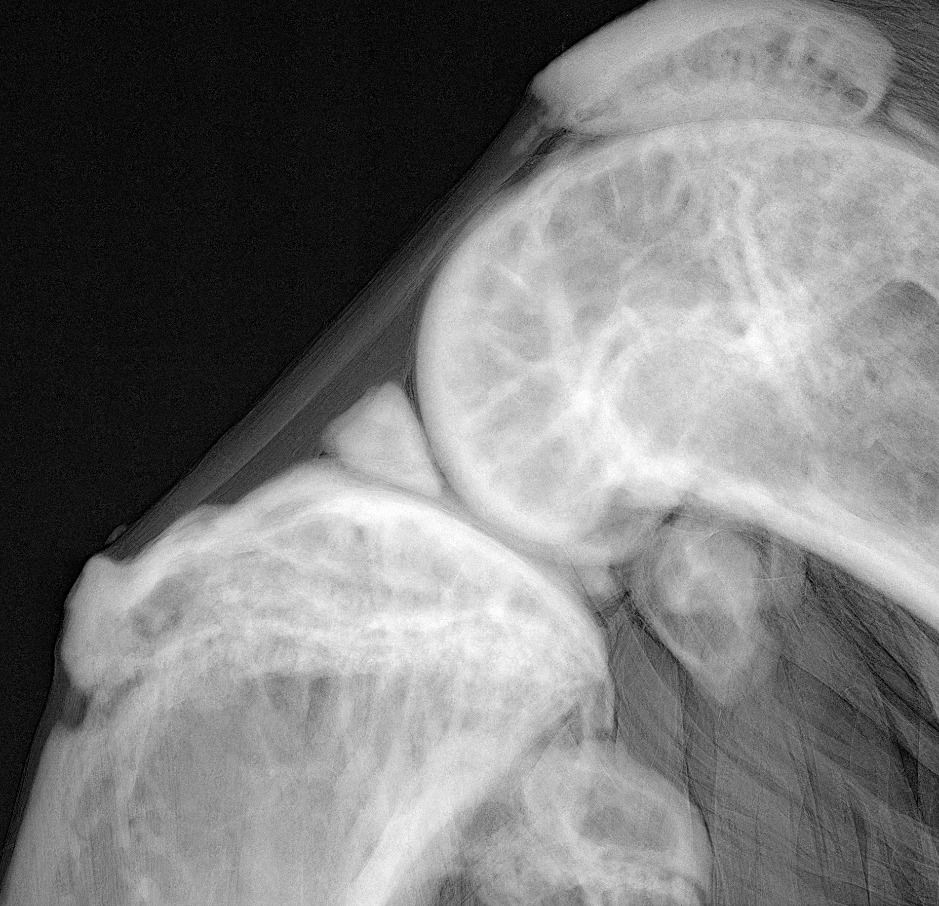
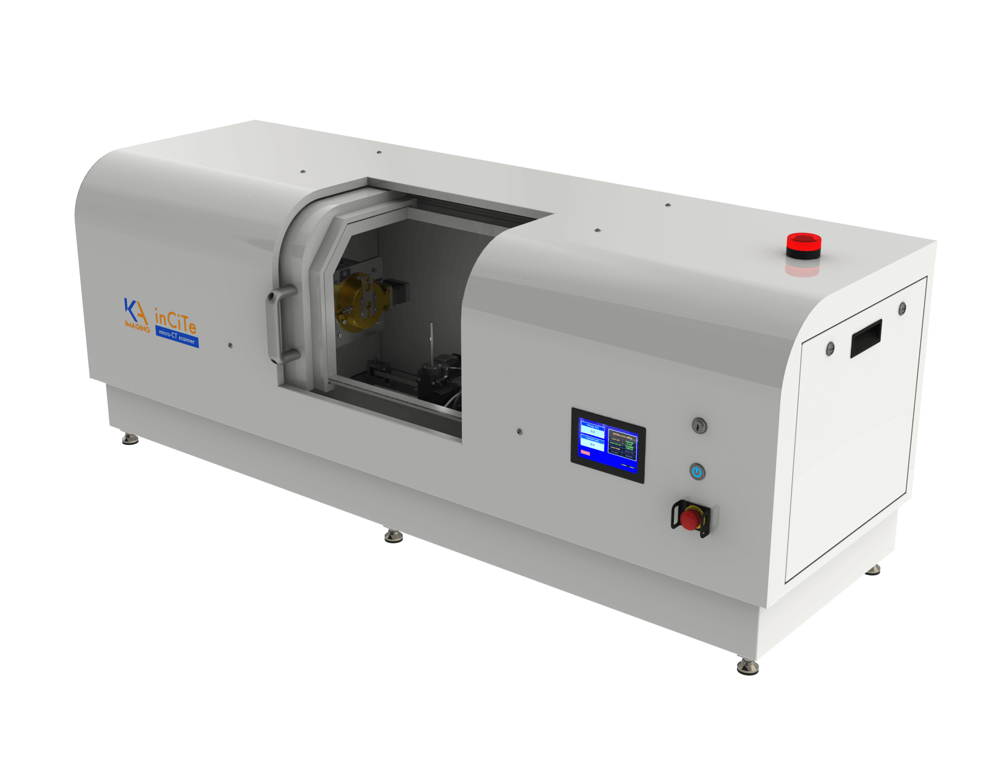
The InCiTe™ 3D X-ray Microscope is a state-of-the-art benchtop micro-CT platform designed to deliver high-resolution, three-dimensional visualization of biological tissues, bone microarchitecture, and advanced materials. Its phase contrast imaging capabilities reveal fine structural details in trabecular and cortical bone, implant-tissue interfaces, and low-density specimens difficult to resolve using conventional X-ray techniques. Compact yet powerful, the InCiTe™ 3D delivers exceptional insights, revolutionizing applications in preclinical orthopaedic research.
Our imaging agents provide a comprehensive portfolio of dedicated, high-quality contrast agents developed specifically for preclinical research. We offer a versatile range of contrast agents designed to support all major imaging modalities — MRI, CT, ultrasound, and optical imaging.