System Used:
Prospect T1
New Model Available

Fangming Wang, Xuejiao Dong, Jing Wang, Feiya Yang, Donghua Liu, Jianlin Ma, Shuai Liu, Dehua Chang, & Nianzeng Xing
Prospect T1 System Publication Highlight
Summary
Using natural killer (NK) cell therapy, researchers recently showed therapeutic efficacy against castration-resistant prostate cancer (CRPC)1. CRPC is an advanced form of prostate cancer with a poor prognosis, an impaired quality of life, and no current known cure2,3. Prostate cancer typically needs testosterone to grow, but CRPC continues growing even when testosterone levels are low, which limits treatments. Using the Prospect T1 high-frequency ultrasound system (now offering the new model, Prospect T2), this group showed that treatment with NK cells significantly reduced both the overall subcutaneous tumor size as well as the blood supply within that tumor – overall showing that NK cell treatment could be a potential therapeutic avenue for CRPC.
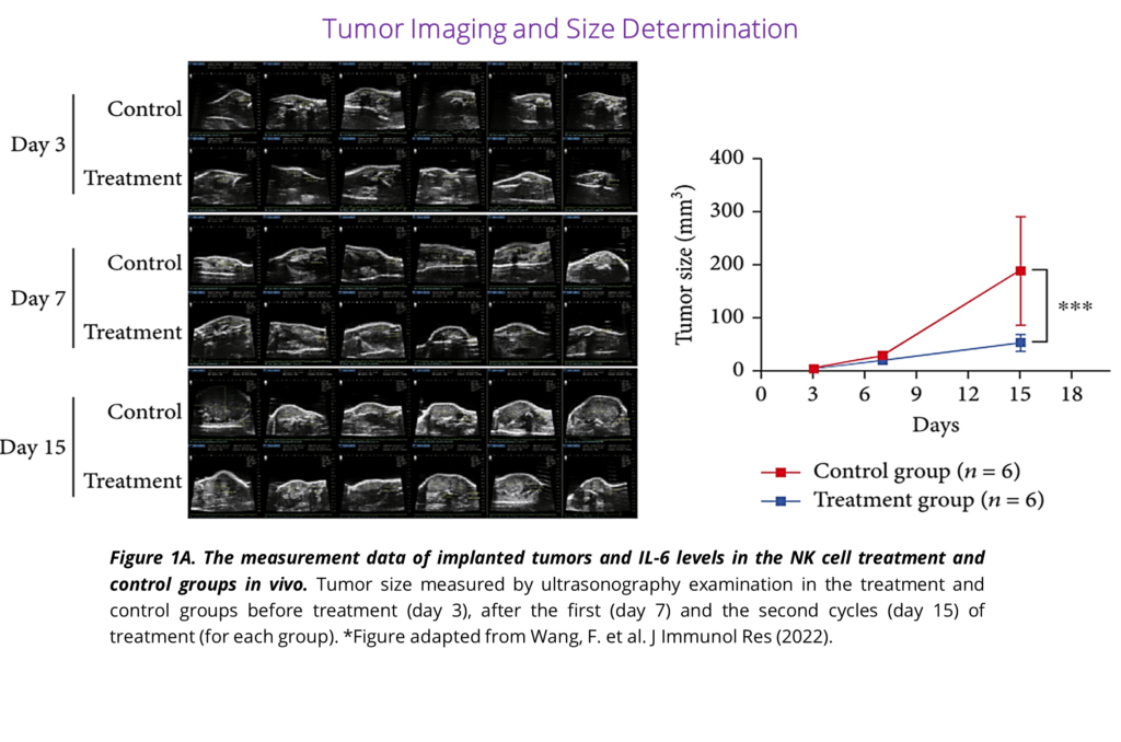
This group estimated tumor size with both calipers and 2D ultrasound imaging, with the ultrasound imaging highlighted in Figure 1A showing the NK treatment group had reduced tumor growth curves in their xenograft mouse model. Using calipers, although common, comes with its’ known biases – mainly, differences in tumor compressibility, surrounding fat, and irregular tumor shapes as well as higher interoperator variability, all of which will affect the accuracy of this measurement4; measurements from ultrasound offer a more reliable method for obtaining accurate tumor sizes and growths5.
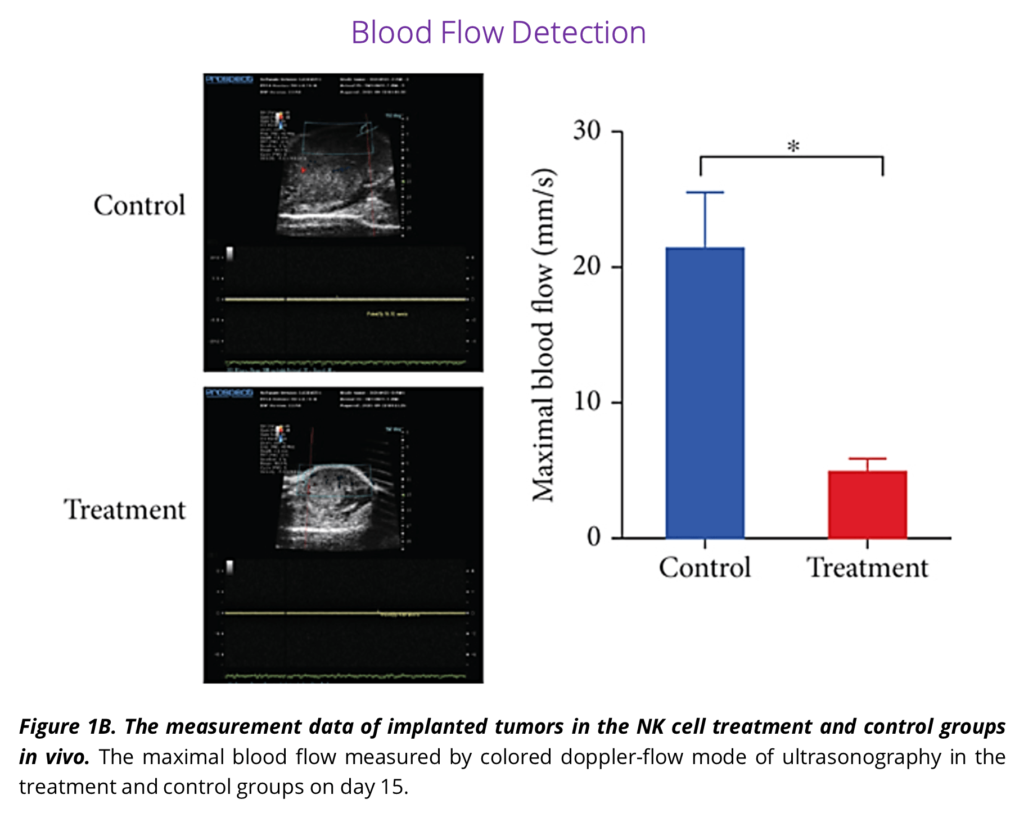
This group also analyzed maximal blood flow within the tumors using ultrasound imaging, as highlighted in Figure 1B showing the NK treatment group has less vascularized tumors than the controls. Blood flow within a tumor can be a crucial measure since blood vessels are essential for maintaining tumor growth, drug responsiveness, as well as playing a role in future metastasis6.
The Prospect T2 is a high-frequency (20-50MHz) and high-resolution (~30μm) ultrasound imaging system. Although subcutaneous tumors were imaged in this work, the system can image various tumor types, such as cell or patient-derived xenografts, transgenic, orthotopic, or other types of models. Tumors are reliably and accurately detectable as small as ~1mm and can be measured with 2D linear and 3D volumetric measurements. Color, power, and pulse-wave Doppler are included with the Prospect T2 and are common ways to measure blood flow within tumors. If microvascularity within tumors is of interest, contrast imaging with microbubbles can also be imaged. Check out the Prospect T2 High-Frequency Ultrasound System website page below – if you would like to discuss your research needs or to request a demonstration of the system with your specific models.
References:
1. Wang, F. et al. Allogeneic Expanded Human Peripheral NK Cells Control Prostate Cancer Growth in a Preclinical Mouse Model of Castration-Resistant Prostate Cancer. J Immunol Res 2022, (2022).
2. Treatments for castration-resistant prostate cancer | Canadian Cancer Society. https://cancer.ca/en/cancer-information/cancer-types/prostate/treatment/castration-resistant-prostate-cancer.
3. ČAPOUN, O. et al. Prognosis of Castration-resistant Prostate Cancer Patients – Use of the AdnaTest® System for Detection of Circulating Tumor Cells. Anticancer Res 36, (2016).
4. Kersemans, V., Cornelissen, B., Allen, P. D., Beech, J. S. & Smart, S. C. Subcutaneous tumor volume measurement in the awake, manually restrained mouse using MRI. Journal of Magnetic Resonance Imaging 37, 1499–1504 (2013).
5. Ayers, G. D. et al. Volume of preclinical xenograft tumors is more accurately assessed by ultrasound imaging than manual caliper measurements. J Ultrasound Med 29, 891 (2010).
6. Zhang, K. & Waxman, D. J. Impact of Tumor Vascularity on Responsiveness to Anti-angiogenesis in a Prostate Cancer Stem Cell-derived Tumor Model. Mol Cancer Ther 12, 787–798 (2013)