(April 9th, 2026) Webinar: Driving NAMs-Aligned Preclinical Research with the Newton 7.0 Optical Imaging Systems
(April 9th, 2026) Webinar: Driving NAMs-Aligned Preclinical Research with the Newton 7.0 Optical
In this webinar, we will review common preclinical cancer models, including orthotopic and heterotopic tumors in syngeneic and xenogeneic rodents. Further, we will review the traditional measurement strategies for describing cancer outcomes as well as modern imaging modalities.
This webinar will serve as a brief introduction to cancer models and imaging techniques for those who are unfamiliar with preclinical imaging instruments. Further, the webinar will provide in-depth information for users who are interested in learning new measurement techniques to augment their cancer studies. Participants are encouraged to arrive with specific questions, imaging challenges, and new applications they are seeking assistance with.
Overview:
Modern cancer studies have evolved significantly in recent decades. As our understanding of oncology has advanced, so too have our models for studying this umbrella of diseases. Cancers represent a heterogenous cascade of diseases with highly variable aetiology, including originating tissues, genetic mutation makeup, and microenvironment characteristics. Cancer studies demand highly reproducible data that is relevant to what is observed in the clinic. Consequently, great pressure has been placed on developing novel models to more accurately reproduce cancers observed in the clinic.
The most basic cancer models involve studying novel treatments in vitro for direct treatment efficacy before transitioning to rodent models. Understanding how cancers grow in vivo is of critical importance, as they more accurately replicate tumor microenvironments that are observed in the clinic.
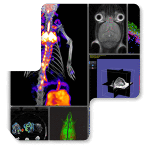
A common hurdle for researchers is being able to accurately and efficiently measure tumor growth or shrinkage in longitudinal research designs. Subcutaneous tumors can be measured manually using calipers, but they are limited by formulaic assumptions based on length and width without considering depth or shape. For manual assessment, internal tumors must be measured ex vivo and are therefore unsuitable for longitudinal studies.
Alternative measurement tools have been developed to overcome these obstacles, including fluorescent, bioluminescent, and 3D ultrasound imaging technologies.
 In this webinar, we will be discussing common methods for studying tumors and how modern imaging technologies can improve your research outcomes.
Learning Objectives:

Dr. James Vanhie holds a Ph.D. from the University of Ottawa in Health Sciences. He previously obtained a Master of Science degree in Kinesiology from Western University and a Bachelor of Science degree in Human Kinetics from the University of Guelph. Throughout 10 years of research experience, Dr. Vanhie has improved our understanding of physiology and disease development across a wide range of fields, including neuromuscular and cardiorespiratory physiology, cancer biology, immune cell development and regulation, and viral oncology. In his present role as a Preclinical Application Specialist, Dr. Vanhie works closely with Scintica’s Prospect T2 High-Frequency Ultrasound imaging system and specializes in providing innovative research solutions for scientists.
(April 9th, 2026) Webinar: Driving NAMs-Aligned Preclinical Research with the Newton 7.0 Optical
(March 12, 2026) Webinar: Preclinical Imaging of Multiple Myeloma Bone Disease by Dual-Energy
(February 25, 2026) Webinar: How to Optimize Your Western Blot Workflow and Choose
(February 12, 2026) Webinar: Sharper Images, Stronger Studies- Optimizing Your Preclinical Ultrasound Techniques
(July 2, 2025) The World’s Smallest Pacemaker: Design and Applications Minimally Invasive. Wireless.
(June 12, 2025) From Scan to Results in Minutes: A Live Demo of