

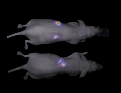


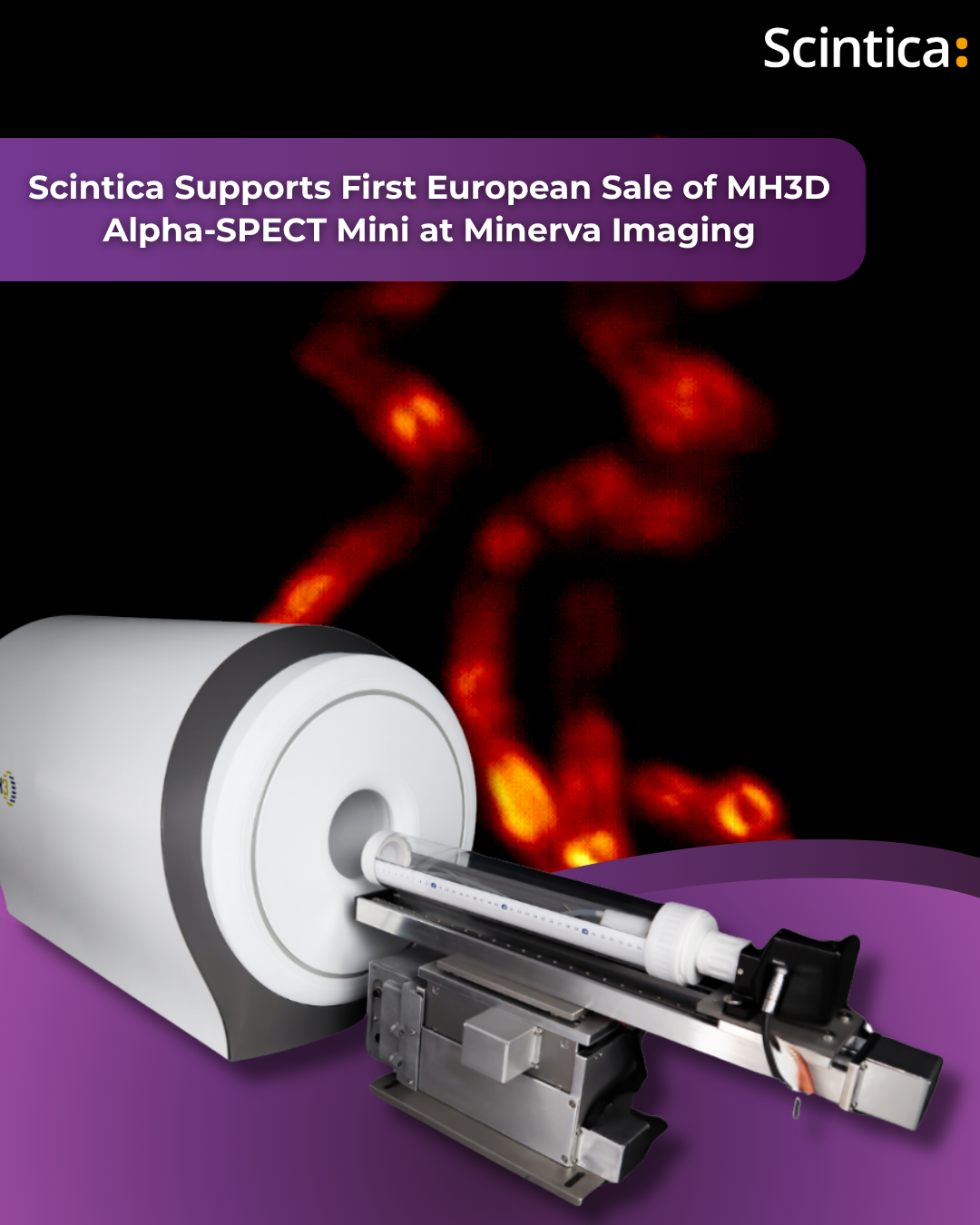
Scintica Supports First European Sale of MH3D Alpha-SPECT™ Mini at Minerva Imaging
London, Canada – Scintica is pleased to announce its role in supporting the
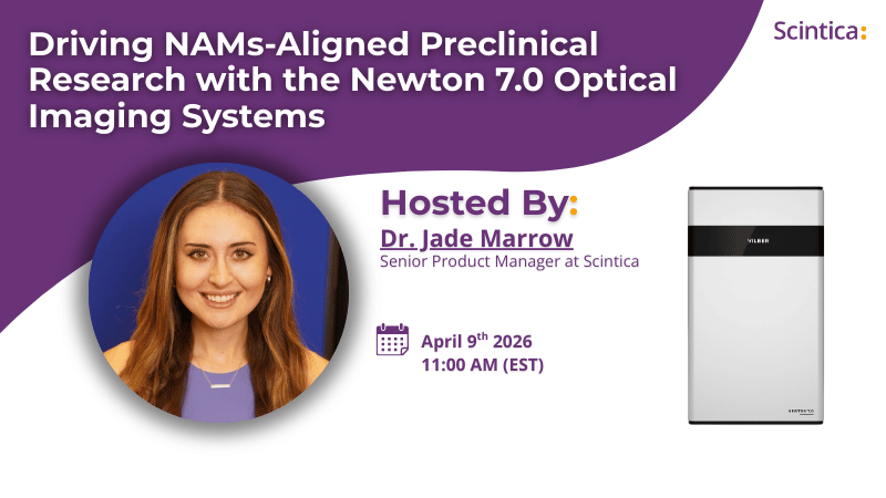
(April 9th, 2026) Webinar: Driving NAMs-Aligned Preclinical Research with the Newton 7.0 Optical Imaging Systems
(April 9th, 2026) Webinar: Driving NAMs-Aligned Preclinical Research with the Newton 7.0 Optical
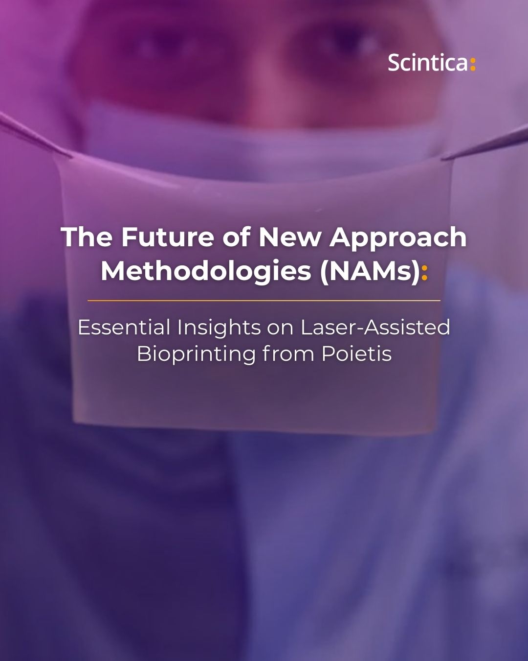
The Future of New Approach Methodologies (NAMs): Essential Insights on Laser-Assisted Bioprinting
As the push for New Approach Methodologies (NAMs) accelerates worldwide, pharmaceutical laboratories and