This study uses longitudinal intravital confocal microscopy in a live PDAC mouse model to show that tumor vasculature is highly heterogeneous and dynamically unstable, with rapidly shifting vessel structure and perfusion that may explain the limited efficacy of anti-angiogenic therapies.
Overview
Pancreatic ductal adenocarcinoma (PDAC) carries a 5-year survival rate below 10%, partly because its vascular biology remains poorly understood. Anti-angiogenic therapies that succeed in colorectal, renal, and lung cancers have consistently failed in PDAC, suggesting the field is working with an incomplete picture.
This study addresses that gap by watching the tumor vasculature evolve in real time, in a living animal. Using a custom pancreatic imaging window implanted in an orthotopic mouse model, the authors performed longitudinal intravital confocal microscopy of the same tumor regions daily for up to 18 days, tracking both vessel structure via fluorescent anti-CD31 antibody labeling and active perfusion via fluorescently labeled red blood cells (RBCs).
Key Findings
- Microscale vascular heterogeneity: Neighboring vessel regions just a few hundred microns apart displayed completely different remodeling behavior — active angiogenesis in one area, dilation and regression in another, simultaneously and within the same tumor.
- Dynamic perfusion instability: Real-time RBC tracking revealed that individual vessel branches oscillated between high flow, near-zero flow, partial recovery, and eventual collapse, suggesting functional perfusion loss likely precedes and drives structural vessel regression.
- A possible explanation for anti-angiogenic trial failures: The spatial and temporal heterogeneity of PDAC vasculature observed here may explain why therapies targeting a single angiogenic pathway (e.g., bevacizumab vs. VEGF) fail to produce consistent clinical benefit , different tumor regions are in different vascular states at any given moment.
- New imaging window design: The team developed a modified stainless-steel pancreatic imaging window with structural gaps to accommodate tumor expansion without causing bleeding , overcoming a critical limitation of earlier designs.
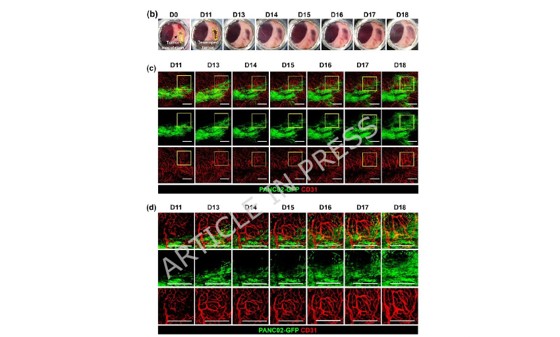
Longitudinal Intravital Imaging of PDAC Tumorigenesis and Vascular Alteration
Longitudinal intravital imaging of Panc02-GFP tumors (green) and CD31+ vasculature (red) from post-modeling day 11 to day 18. Upper panels show wide-field views; lower panels show magnified regions of interest revealing progressive vessel dilation, branching changes, and tumor cell invasion. Scale bar, 500 µm. Imaging acquired using the IVM-CMS system (IVIM Technology, Korea) in confocal mode. Scale bar, 500 μm.
System Used
System: IVM-CMS (IVIM Technology, Korea), commercial intravital confocal and two-photon microscopy platform
Imaging mode: Confocal (all images and videos in this study)
RBC video acquisition: 60-second recordings at 15 fps per session
Longitudinal span: Up to 18 days; 1–2 hours per imaging session
Why This Matters for Research
This paper demonstrates what separates intravital microscopy from all other preclinical imaging modalities: the ability to return to the same tissue, the same vessels, and the same cells across time , in a living animal. Static histological endpoints, the workhorse of preclinical oncology, would have produced averaged snapshots of an inherently dynamic process.
For researchers working in tumor vascular biology, drug delivery, or vascular normalization, this study shows that spatial resolution alone is insufficient. Temporal resolution, returning to the same region of interest across days, is what transforms a microscope into a discovery platform.
Intravital Microscopy
Learn more about the system featured in this article.
Get a Quote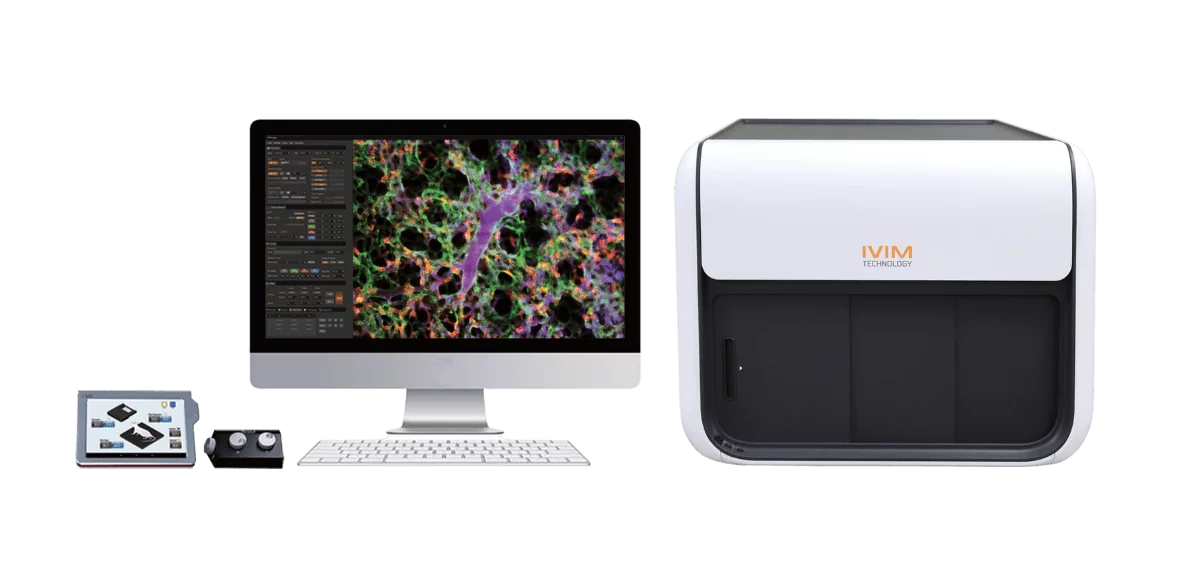
References
Lucia S.E., Hong S., Abdi R. & Kim P. • Cancer Cell International (2026)
• DOI: 10.1186/s12935-026-04363-7