System Used:
NGB-R
M-Series
Organs-on-chips (OoCs) are microengineered systems intended to emulate specific organs or processes of the human body. As such, they show great promise for translational research, as in theory, an OoC would be an in vitro model more directly mimicking human biology than traditional in vivo animal models. Typically, OoCs consist of a small model comprised of perfused channels and cell-laden culture chambers, often featuring tissue interfaces between tissue types, organs, and barrier interfaces. Applications can include drug discovery, toxicity testing, molecular biology, and personalized medicine. In a recent publication by Bosmans et al., the authors review and discuss the future of OoCs, how bioprinting plays a large role, and how single-cell bioprinting through laser-assisted bioprinting will be key to these advances1.
Organs-on-Chip
Akin to a more complex version of a Transwell, OoCs are basically tiny cell culture models but with complex microfluidic structures, designed to replicate the function of a specific organ or process. An OoC can be as small as a thumb, or as large as a microplate, and is typically made on an optically transparent material such as glass, plastic, or polydimethylsiloxane (PDMS). While many approaches have been taken to create OoCs, they are all typically seeded with cells or contain a tissue-engineered construct and are perfused in a way to replicate physiological stresses, all to study cell-cell and cell-ECM interactions. Another use of OoCs includes adding semi-permeable membranes to study systems like the blood-brain barrier or the lungs. Finally, OoCs can also be combined to create an entire interconnected system.
Microarchitecture
he microarchitecture of an OoC, defined as the design and layout of the minute structures and arrangements, has been shown to be key to properly replicate physiologically relevant applications. This has been shown in both a virus infection model and a trabecular bone model, where in each case, the 3D design with proper microarchitecture was able to better mimic desired conditions than 2D or unstructured 3D OoCs. Changes in this microarchitecture are also prevalent in disease state, so it is important to have the ability to modify the architecture instead of using earlier unstructured approaches to OoCs. For this, 3D bioprinting has the potential to become the tool of choice to allow rapid prototyping of intricate designs.
3D Bioprinting Technologies
The 3D bioprinting market has exploded in recent years with numerous technologies used (Figure 1). Broadly, they can be categorized as nozzle-based or light-based, with several sub-categories. The two most common technologies are inkjet drop on demand/micro-valve and bio-extrusion methods. While capable, these are limited in how fast they can print, and can affect cells passing through due to the shear stress they are exposed to. This can lead to lower cell viability and modified morphology. Light-based technologies can overcome these limitations as they work on completely different principles. For example, using the laser-induced forward transfer (LIFT) principle, small micropatterns can be printed extremely rapidly without inducing DNA damage, heat-shock, or applying shear stress to individual cells. With LIFT, a donor slide coated with the material to be transferred is used. A high-power pulsed laser is used to eject tiny amounts off the slide, transferring it to the print substrate.
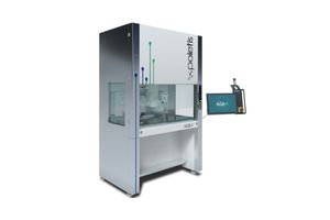
Next Generation Bioprinter (NGB)-R
These features make LIFT, or more broadly, laser-assisted bioprinting (LAB), the optimal choice for developing single-cell (resolution) bioprinting for even denser microprinting of tissues, liquids, and other biomaterials necessary to mimic the microenvironment of actual tissues (Figure 1). The Next Generation Bioprinter (NGB)-R 4D bioprinter is one of, if not the only commercial LAB-based printers on the market, here to take your research to new heights. In addition, the NGB-R also includes inkjet drop on demand and extrusion bioprinting technologies, allowing research groups already using bioprinting to transfer their existing designs over, while adding the capabilities of LAB. These are all combined with a 6 degree-of-freedom robotic arm and integrated microscope to ensure the highest precision for replicating the complex tissue structures needed for advanced disease modeling, drug discovery, and personalized medicine.

Figure 1: LIFT mechanism and notable patterning examples. Reproduced from Bosmans et al. (2023).
References
- Bosmans, C. et al. Towards single-cell bioprinting: micropatterning tools for organ-on-chip development. (2023) doi:10.1016/j.tibtech.2023.11.014.