M-Series
Application Note:
Deep Brain Imaging Utilizing Novel Surgical Techniques: Cranial Imaging
Window and Cannula with Intravital Tunable Two-Photon Microscopy
Intravital brain imaging is a powerful technique for observing neurons, glial cells, and their surrounding environment in living animals. It enables the study of neurological diseases, drug efficacy, and the immune system. Two-photon microscopy, in particular, is an essential tool for in vivo imaging. It employs two lower-energy photons to excite fluorophores, which allows for deep tissue imaging due to the reduced scattering of longer-wavelength infrared light. This results in clearer images with minimal background noise from out-of-focus light. The use of tunable two-photon lasers further enhances imaging by adjusting excitation wavelengths, making it possible to efficiently excite a broader range of fluorophores, allowing for more versatile and comprehensive imaging across various biological contexts.
Traditionally, the cranial imaging window (CIW) has been employed for brain imaging. This method involves creating a hole in the skull and placing a transparent window over the exposed brain tissue, facilitating observation via microscopy. However, in vivo brain imaging has been primarily restricted to cortical regions, with significant challenges in visualizing deeper brain structures, such as the hippocampus. These limitations impede our ability to thoroughly investigate the complex dynamics of deeper brain regions in living organisms.
The Cranial Imaging Cannula (CIC) is a novel surgical technique that enables real-time imaging of deep brain areas, including the hippocampus and hypothalamus—regions critical for studying neurodegenerative diseases and neural dynamics. By precisely inserting a cannula into the brain, the CIC allows for long-term observation of deep brain structures with minimal tissue damage, making it ideal for longitudinal studies. This technique can be used to track drug effects or disease progression over extended periods, such as weeks or months.
In our study, we first utilized tunable two-photon microscopy in combination with the CIW technique to observe neuronal activity in Thy1-GCaMP6f transgenic mice. GCaMP6f is the fastest calcium indicator for detecting cytoplasmic free calcium. In these mice, GCaMP6f is expressed under the Thy1 promoter, which enables the detection of neuronal activity through calcium fluctuations corresponding to neuronal firing. This setup allowed us to visualizeGCaMP6f fluorescence in cortical neurons.
Additionally, we employed a fluorophore-conjugated CD31 antibody (CD31-Setau647) for in vivo blood vessel labeling. Using the tunable two-photon laser, we optimized the excitation wavelength to simultaneously observe both GCaMP6f and Setau647 signals.
Next, we explored the depth of brain visualization achievable using the CIW technique combined with two-photon microscopy, comparing it to the CIC method for accessing deeper brain regions. This comparison allowed us to assess the effectiveness of each technique, particularly for visualizing deep brain areas.
Materials & Methods
Mouse Models
Thy1-GCaMP6f transgenic mice were used to monitor neuronal activity. These mice are genetically modified to express GCaMP6f under the Thy1 promoter, allowing for stable and reproducible fluorescence in neurons. To visualize cortical neurons, Thy1-M transgenic mice were used. For imaging the hippocampal CA1 region, C57BL/6N mice were selected.
Cranial Imaging Window (CIW)
For long-term and stable imaging of the brain cortex, the Cranial Imaging Window (CIW)technique was employed. Mice were anesthetized using a mixture of Zoletil, Rumpun, and saline, administered via intramuscular injection to ensure proper sedation and pain management. After anesthesia, the mice were positioned in a stereotaxic frame to precisely target the cortex. A craniotomy was performed, followed by placing a cover glass over the exposed brain and securing it with optical bond and dental cement.
Cranial Imaging Cannula (CIC) Surgery
After anesthesia, mice were placed in a stereotaxic frame and 3mm diameter hole was made on the skull with the same protocol with CIW surgery for precise targeting of the hippocampal CA1 region. After exposing the brain through a cranial opening, the meninges and part of the cortex are removed using suction to reveal the hippocampus. Once the bleeding has stopped, a cranial imaging cannula is inserted and covered with a 3mm cover glass. The incision area is then secured using fast-curing adhesive and dental acrylic resin except for the cannula.
Blood Vessel Labelling
To visualize blood vessels in live mice, fluorophore-conjugated antibodies (anti-CD31-Setau647, IVIM Technology) were administered via intravenous injection. Two hours post-injection, intravital confocal and two-photon microscopy (IVM-CM3) were used to capture blood vessel images.
Two-Photon Imaging
After the surgical procedure, mice were placed in cage to recovery and observed for at least2~4 weeks under inflammation control with Rimadyl injection(Carprofen, zoetis).
For visualizing the blood vessels, IVI Tag TM In Vivo Labeling Kit (IVIM Technology, Korea Republic) was utilized. Under the anesthesia, 25 μL of Kit for labeling the endothelial cells was injected intravenously through tail vein route. After 1 hour, the mouse was mounted on the stereotaxic stage with heating function in the IVM-CM3 intravital microscope (IVIM Technology, Korea Republic).
Mice were placed in a stereotaxic frame for stability during imaging. Two-photon imaging was performed using intravital confocal and two-photon microscopy (IVM-CM3).


Figure 1: Mouse animal models implanted with (A) Cranial imaging window, and (B) Cranial imaging cannula.
Results

Blood Vessel GCamPf6
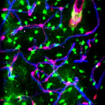
Figure 2: Visualization of GCaMP Expression

Figure 3: Two-Photon Brain Imaging in CIW-Implanted Thy1-M-GFP Mice.
Thy1-GCaMP6f mice express endogenous GFP signal for detecting Ca2+ ion dependent neuronal activities. If neurons get to be activated, GCaMP6f bound to calcium ions rapidly expresses its endogenous GFP, which make it advantage for studying neuronal cell activities. For visualizing the real-time changes of neuronal activity, we used this mouse species after CIW implantation.
Our data (Fig1A and 2A) showed that some neurons had increased GFP, referring that we could acquire the real-time cellular dynamics in brain with CIW implanted mouse model.
In Thy1-M-GFP mice implanted with a cranial imaging window (CIW), we measured the brain depth visualized using a 920 nm two-photon laser. Second harmonic generation (SHG)signals from collagen indicated its presence in the superficial layers. Blood vessels were observed below the collagen layer, with larger vessels present in the upper regions and micro-vessels becoming predominant at greater depths. Neurons labeled with Thy1-M-GFPwere visualized, demonstrating that the two-photon microscopy combined with CIW technology enabled us to observe up to approximately 600 μm within the cortical areaInThy1-M-GFP mice implanted with a cranial imaging window (CIW), we measured the brain depth visualized using a 920 nm two-photon laser. Second harmonic generation (SHG)signals from collagen indicated its presence in the superficial layers. Blood vessels were observed below the collagen layer, with larger vessels present in the upper regions and micro-vessels becoming predominant at greater depths. Neurons labeled with Thy1-M-GFPwere visualized, demonstrating that the two-photon microscopy combined with CIW technology enabled us to observe up to approximately 600 μm within the cortical area.

Blood vessel GCamPf6
Figure 4: Two-Photon Hippocampal CA1 Imaging in CIC-Implanted Mice
To overcome limitations in imaging depth and visualize deep brain regions, we implanted a cranial imaging cannula (CIC) in mice. Using a 920 nm two-photon laser, we successfully observed blood vessels in the hippocampal CA1 region in CIC-implanted mice. These findings confirmed that the CIC technique allows for real-time imaging of deep brain areas, such as the hippocampus, in living mice. This advancement extends our ability to observe not only cortical neuronal activity and blood vessels but also deeper brain regions like the hippocampus, providing a crucial tool for studying neurological diseases and drug responses in previously inaccessible brain areas.
References
Dana, Hod, et al. “Thy1-GCaMP6 transgenic mice for neuronal population imaging in vivo.” PloS one9 (2014): e108697. https://doi.org/10.1371/journal.pone.0108697
Bradley, John E., Gustavo Ramirez, and James S. Hagood. “Roles and regulation of Thy‐1, a context‐dependent modulator of cell phenotype.”Biofactors3 (2009): 258-265.https://doi.org/10.1002/biof.41
Benninger, Richard KP, and David W. Piston. “Two‐photon excitation microscopy for the study of living cells and tissues.” Current protocols in cell biology1 (2013): 4-11.https://doi.org/10.1002%2F0471143030.cb0411s59
2. 3. 1.
Want to learn more about the Intravital Microscopy (IVM)?