Expanding the Possibilities of Preclinical Research with the Newton Optical Imaging System
Understanding biological processes as they occur in living organisms is essential for advancing translational research. Whether investigating cancer progression, infectious disease, immune cell trafficking, vascular function, drug delivery, or neurology, researchers rely on non-invasive imaging technologies that provide real-time biological insights while minimizing animal use.
The Newton optical imaging platform is designed to address this need through highly sensitive bioluminescence, fluorescence, and near-infrared imaging. By supporting imaging across visible (VIS), near-infrared (NIR-I), and second near-infrared (NIR-II) wavelengths, the system enables researchers to visualize molecular and cellular events deep within living tissues without exposing subjects to harmful ionizing radiation.

The Value of Optical Imaging
Traditional endpoint analyses often require euthanizing animals at multiple time points, introducing variability in the results and increasing study costs. With optical imaging, the same animal can be repeatedly monitored throughout the entirety of an experiment. This longitudinal capability enhances statistical power, reduces animal numbers, and provides a more complete picture of disease progression and treatment response.
Applications of Optical Imaging with the Newton Systems
1) Oncology
Optical imaging is widely used in cancer research to monitor tumor growth, metastasis, and treatment response. Tumor cells expressing luciferase can be tracked using bioluminescence imaging to quantify viable tumor burden over time. Fluorescent probes help visualize molecular targets, tumor-associated biomarkers, and therapeutic accumulation within tumors.
With the Newton FT-900, NIR-II imaging provides improved image contrast and tissue penetration compared to visible and NIR-I imaging, making it particularly useful for imaging deep-seated tumors and assessing drug delivery to tumor tissue.

2) Cardiovascular Mapping
The enhanced tissue penetration of NIR-II imaging (Newton FT-900) makes it especially useful for vascular applications. Less light scattering and absorption by hemoglobin allows for the visualization of vascular architecture and perfusion. Cardiac and cerebral blood flow patterns and real-time vascular dynamics can be assessed using contrast agents. This is relevant in oncology, cardiovascular research, regenerative medicine, and neuroscience. Researchers can evaluate:
- Tumor angiogenesis
- Vascular remodelling
- Tissue perfusion
- Nanoparticle transport through the vasculature

3) Immunology
Fluorescent and bioluminescent reporters can be used to monitor immune cell migration and localization in vivo. By enabling non-invasive monitoring, optical imaging allows researchers to follow immune responses dynamically throughout the course of an experiment. Specific uses cases include:
- Inflammatory cell recruitment
- T-cell trafficking during immunotherapy (e.g., cell-based therapies, CAR-T cells)

4) Infectious Disease Research
Reporter-labelled pathogens can be tracked to measure the onset of infection, spread, and therapeutic response. With the Newton systems, researchers can non-invasively study infections in living subjects and plants while also evaluating the effectiveness of antimicrobial treatments and therapeutic interventions. Applications include:
- Tracking bacterial or viral infections in live animals and plants
- Evaluating treatment response
- Disease progression monitoring

5) Neurology
In addition to traditional BLI, the growing adoption of NIR-II imaging is creating new opportunities in neurological research. Because longer wavelengths penetrate tissue more effectively, researchers can visualize signals deeper (i.e., in the brain) while still maintaining high image quality.

6) Drug Delivery and Biodistribution
The Newton enables researchers to track fluorescently labeled drugs, antibodies, peptides, and nanoparticles in vivo. These studies help determine whether a therapy has reached its intended target, how long it remains in circulation, and where off-target accumulation occurs. Applications include:
- Evaluating nanoparticle targeting efficiency
- Comparing drug formulations
- Monitoring antibody distribution
- Assessing pharmacokinetics and clearance
The increased penetration depth and reduced tissue autofluorescence of NIR-II imaging in the Newton FT-900 can improve visualization of labeled therapeutics in deeper tissues.

7) Probe Development & Validation
There is an increasing need for commercially available NIR-II fluorophores. The Newton FT-900 enables proper evaluation of probe radiance, target specificity, and metabolism/clearance. This includes organic fluorophores, rare-earth nanoparticles, quantum dots and nanomaterials, small molecule dyes, etc.
8) Ex Vivo Imaging
While the Newton platform is widely used for small animal research, researchers can also image excised organs, microplates, Eppendorf tubes, and other ex vivo samples using the same platform. This flexibility allows investigators to combine whole-animal imaging with downstream tissue analysis within a unified workflow.

Supporting Translational Research
The combination of bioluminescence and fluorescence imaging in VIS, NIR-I, and NIR-II makes the Newton a versatile platform for a broad range of preclinical applications. Common research areas include oncology, immunology, infectious disease, drug development, vascular biology, and nanomedicine. By enabling non-invasive, longitudinal imaging, the system provides researchers with quantitative data on biological processes, therapeutic efficacy, and biodistribution throughout the course of a study.
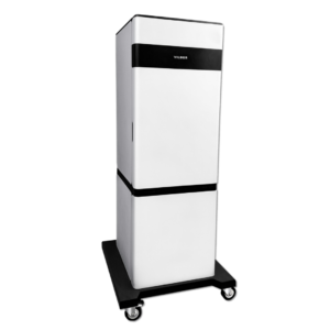
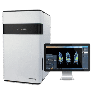
System Models Discussed in This Post
Book a meeting with one of our scientists to learn how the Newton Optical Imaging System can support your research