
Publication Highlight: Target & Shoot Spheroid Bioprinting

Original study: Nilsson Hall G. et al.
Published in: Biofabrication (2024)
DOI: 10.1088/1758-5090/ad6e1a

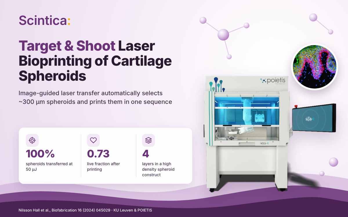
Researchers at KU Leuven and POIETIS combined laser-assisted bioprinting with a custom imaging system to automatically find, target and transfer multiple large cartilaginous spheroids in a single sequence. At 50 µJ, the ‘target and shoot’ system transferred 100% of ~300 µm human periosteum-derived cell (hPDC) spheroids (n = 30), and the team used it to build a four-layered, high-density spheroid construct. 

The challenge: placing living building blocks precisely, and fast enough

Multicellular spheroids are attractive building blocks for bottom-up tissue engineering, but assembling them into designed structures is hard. The authors point to three recurring limits of current approaches: 

  • Extrusion bioprinting: low spheroid density, limited control over where spheroids land, and nozzle clogging. 
  • Aspiration-assisted bioprinting: precise placement, but one spheroid per operation, with several minutes per transfer. 
  • Self-assembly in containers: fusion happens without designed architecture, so final shape can differ from what was intended.
 

Laser-assisted bioprinting (LAB) is nozzle-free, which makes it a candidate to avoid clogging. The open question was whether it could handle large, matrix-rich microtissues rather than single cells, and do so with automated selection. 

How 'target and shoot' works

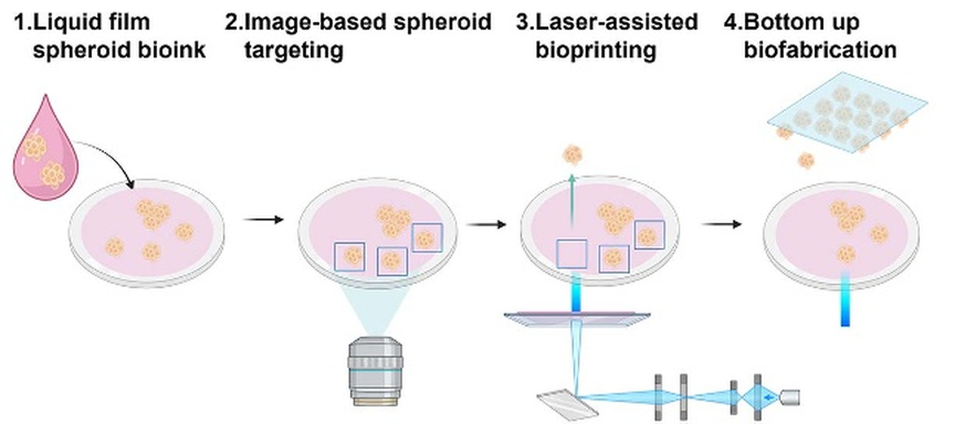
POIETIS developed a custom imaging system and installed it on the LAB workstation; the combination is what the authors call the ‘target and shoot’ system. The workflow runs in four steps: 

Scheme 1. Spheroids in a liquid film are imaged, automatically targeted, transferred by laser, and assembled bottom-up into a predesigned pattern. Source: Nilsson Hall et al., Biofabrication 16 (2024) 045029, CC BY 4.0.

  1. Liquid film loading. Spheroids are suspended in culture medium on the donor. No hydrogel or bioink is used for the transfer itself.

  2. Image-based targeting. The software distinguishes separated spheroids from agglomerates and aims the laser at the geometric center of each selected spheroid.

  3. Laser transfer (LIPMO). For large spheroids, the team developed laser induced propulsion of mesoscopic objects (LIPMO). Energy is deposited just below a single spheroid and propels it ballistically to the receiver, using 8 µl of liquid versus 30 µl for conventional laser-induced forward transfer (LIFT).

  4. Bottom-up assembly. Spheroids are placed in a CAD-defined pattern on the receiver, layer by layer.

Key results

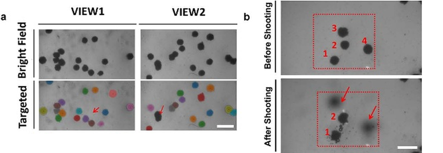
Figure 6(a, b). (a) Automated selection: coloured spheroids are selected and center-targeted; large aggregates are excluded. (b) Spheroids before and after a single laser shoot sequence. Scale bars 500 µm. Source: Nilsson Hall et al., Biofabrication 16 (2024) 045029, CC BY 4.0.

  • Automated selection. The imaging system separated single spheroids from agglomerates and targeted the laser on each selected spheroid’s center.

  • Multiple spheroids per sequence. Two out of four targeted spheroids were transferred with one laser shoot sequence.

  • Energy-dependent transfer efficiency. Transfer rose with laser energy and reached 100% at 50 µJ (n = 30):
Laser energy (µJ) Spheroids transferred
50
100%
40
50%
35
20%
  • Cells survive transfer. Large hPDC spheroids printed by LIPMO at 50 µJ showed a live fraction of 0.73 ± 0.03 (normalized fluorescence).
     
  • Four-layered, high-density construct. Spheroids were laser-printed onto bioextruded collagen type I films, with a 6-axis robotic arm moving the construct between the LAB and bioextrusion work areas. H&E staining confirmed the four-layered structure. 

  • Throughput. Multiple targeted spheroids can be transferred during an 8 ns laser pulse. The authors report an order-of-magnitude gain in process efficiency over aspiration-based methods, which handle one spheroid at a time over several minutes. 

Why it matters

The study moves laser-assisted bioprinting from single cells to matrix-rich, pre-differentiated microtissues, with automated selection built in. For researchers building tissues bottom-up, this opens three practical possibilities:

  • Designed architecture. Spheroids land where the CAD pattern puts them, supporting studies of how spatial arrangement drives fusion and tissue formation. 
  • Quality built into each block. Each spheroid carries its acquired phenotype, an approach the authors link to quality-by-design principles for future regulatory translation. 
  • Mixed niches. Precise placement could combine different spheroid types in one construct, for example, to include vascular structures.
 

The authors position the work as a proof of concept for tissue-engineered implants and organ-on-a-chip models, and name improved spheroid-suspension films and further multi-targeting automation as next steps. 

System Used

NGB-R

3D High-Resolution Multi-Modal Robotic-Assisted Bioprinting System

The NGB-R is a multimodal, 3D bioprinting platform designed and developed to print live tissues and organs. Combining laser-assisted, micro-valve, and extrusion bioprinting, the NGB-R enables true versatility of bioprinting (from cells to spheroids) and offers the possibility of using a large number of biomaterials and hydrogels.

View key capabilities
  • Multi-Modality, Laser-Assisted, Bio-extrusion, and Micro-valve Bioprinting Techniques

  • Robotic-assisted
  • High cellular viability (>95%)
  • Integrated microscopy and image analysis platform
References

Nilsson Hall G, Fan Y, Viellerobe B, Iazzolino A, Dimopoulos A, Poiron C, Clapies A, Luyten F P, Guillemot F and Papantoniou I. Laser-assisted bioprinting of targeted cartilaginous spheroids for high density bottom-up tissue engineering. Biofabrication 16 (2024) 045029. Open access. doi.org/10.1088/1758-5090/ad6e1a

*Information presented on this page and website reflects the data, product specifications, application information, and regulatory status available at the time of publication. Product features, specifications, availability, pricing, and regulatory information may change without notice. Please consult the manufacturer or contact Scintica for the most current information.

*This content is provided for informational purposes only and is based on information available at the time of publication. While reasonable efforts have been made to ensure accuracy, product specifications, performance data, regulatory status, availability, and other details may change without notice. Scintica makes no representations or warranties regarding the ongoing accuracy or completeness of the information presented and recommends verifying all information with the manufacturer before making purchasing or research decisions.