Intravital Microscopy- Real-time Tracking of Osteoclast Activity for Bone Disease Studies
Dynamic Microenvironment of Bone
Bone maintains a dynamic balance between formation through osteoblasts and resorption through osteoclasts. Disruption of this balance, particularly through excessive osteoclast activity, is central to the development of several bone diseases, including osteoporosis, arthritis, and bone metastasis. Increased bone resorption can lead to significant clinical challenges, such as an increased risk of fractures and immobility, contributing to higher mortality rates.
Osteoclast Activity Modulation
Osteoclasts, which are formed from monocytes, resorb bone by secreting hydrochloric acid, proteases like cathepsin K (CatK), and matrix metalloproteinases (MMPs). Current treatments for osteolysis aim to suppress osteoclast activity but are often associated with adverse effects, such as fractures and jaw necrosis. Additionally, these therapies do not effectively prolong survival in cancer patients with bone metastasis. A key limitation in osteoclast-targeted therapy development is the lack of real-time in vivo monitoring of osteoclast number and activity, as traditional histological methods provide only static, two-dimensional snapshots of osteoclast behavior. To address this gap, recent advancements in molecular optical imaging, particularly fluorogenic probes for CatK, have enabled real-time monitoring of osteoclast activity in living animals.
Intravital Microscopy for Real-time High-Resolution Imaging
Intravital microscopy is a sophisticated imaging technique that enables real-time visualization of biological processes within living organisms. While macroscopic imaging methods provide valuable insights, they typically offer only static snapshots of biological phenomena at a larger scale, often with limited resolution. Intravital microscopy delivers dynamic, in vivo insights, capturing complex physiological events as they unfold within their natural context. This provides a more accurate representation of biological conditions compared to static, ex vivo studies by allowing researchers to observe cellular behaviors, tissue interactions, and physiological responses over extended periods, all within the living organism’s natural environment. Additionally, the use of various conjugated antibody labels facilitates the visualization of multiple cells and structural features, further enhancing the depth of analysis. Ultimately, intravital microscopy serves as an invaluable tool, offering access to the dynamic nature of living systems in ways that other imaging modalities cannot replicate (Fig 1).


Figure 1: Intravital imaging of cranial bone marrow (Left) allows for easy monitoring of transplanted cells in specific spots (Right).
Intravital Microscopy is an advanced imaging technique that enables real-time visualization of osteoclast activity in living organisms, surpassing traditional histology in understanding bone disease.
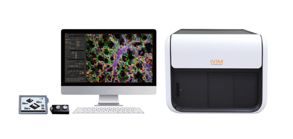
The IVM System: IVIM Technology
IVIM Technology has meticulously designed a system that not only advances imaging technology but also prioritizes user experience. The IVM system is an all-in-one box solution that seamlessly integrates all essential features required for hands-free imaging at high speed and resolution. Every aspect of the system has been meticulously engineered to ensure maximum comfort and efficiency for researchers.
The system is equipped with a range of features, including integrated apparatus for heating, anesthesia administration, window chamber observation, and motion compensation function for long-term monitoring of the tissues and organs. These components work harmoniously to maintain optimal physiological conditions. The system has a compact footprint without compromising performance.
Whether utilizing confocal or two-photon imaging or both, the system boasts the smallest footprint on the market. This not only optimizes laboratory space but also enhances accessibility, giving the researchers the freedom to place the system in any lab space.
Label free Imaging of Collagen Structure Using Second Harmonic Generation (SHG)
With the two-photon modality, the user can utilize second harmonic generation (SHG), which is a label free imaging process capable of detecting striated muscle or collagen networks (Fig 2). SHG imaging has been vital for bone studies as changes in collagen network structure in various bone disease models are easily observed.

Figure 2: SHG imaging of skeletal muscle to help visual neuromuscular junction.
Tracking Osteoclast Activation in Real-Time
This study utilizes high-speed imaging capabilities of IVM to observe osteoclast activity within their natural bone disease context, offering valuable insights into bone health. The study uses a Cathepsin K (CatK)-responsive fluorescent probe (CatKP1) to track osteoclast activity in three different mouse models of bone disease including osteoclastogenesis, osteoporosis, and breast cancer bone metastasis. The probe exhibited a strong fluorescence response when active CatK, an enzyme linked to osteoclast function, was present (Fig 3).
Through this technique, the researchers were able to monitor increases in osteoclast activity in the hindlimb long bones and observe its reduction following pharmacological intervention, all without needing to sacrifice the animals. Two-photon intravital imaging allowed for the observation of osteoclasts at single-cell resolution, demonstrating that CatKP1 is a powerful tool for investigating osteoclast behavior in disease and could be useful for developing new therapeutic strategies.

Blood vessel GCamPf6
Figure 4: Two-Photon Hippocampal CA1 Imaging in CIC-Implanted Mice
References
Koo S, Lee EJ, Xiong H, Yun DH, McDonald MM, Park SI, Kim JS. Real-Time Live Imaging of Osteoclast Activation via Cathepsin K Activity in Bone Diseases. Angew Chem Int Ed Engl. 2024 Feb 5;63(6):e202318459. doi: 10.1002/anie.202318459. Epub 2023 Dec 29. PMID: 3810541
Want to learn more about the Intravital Microscopy (IVM)?