
Ahn, S., Yoon, J.-Y., & Kim, P. (2024).
System Used:
Intravital Microscopy
Overview:
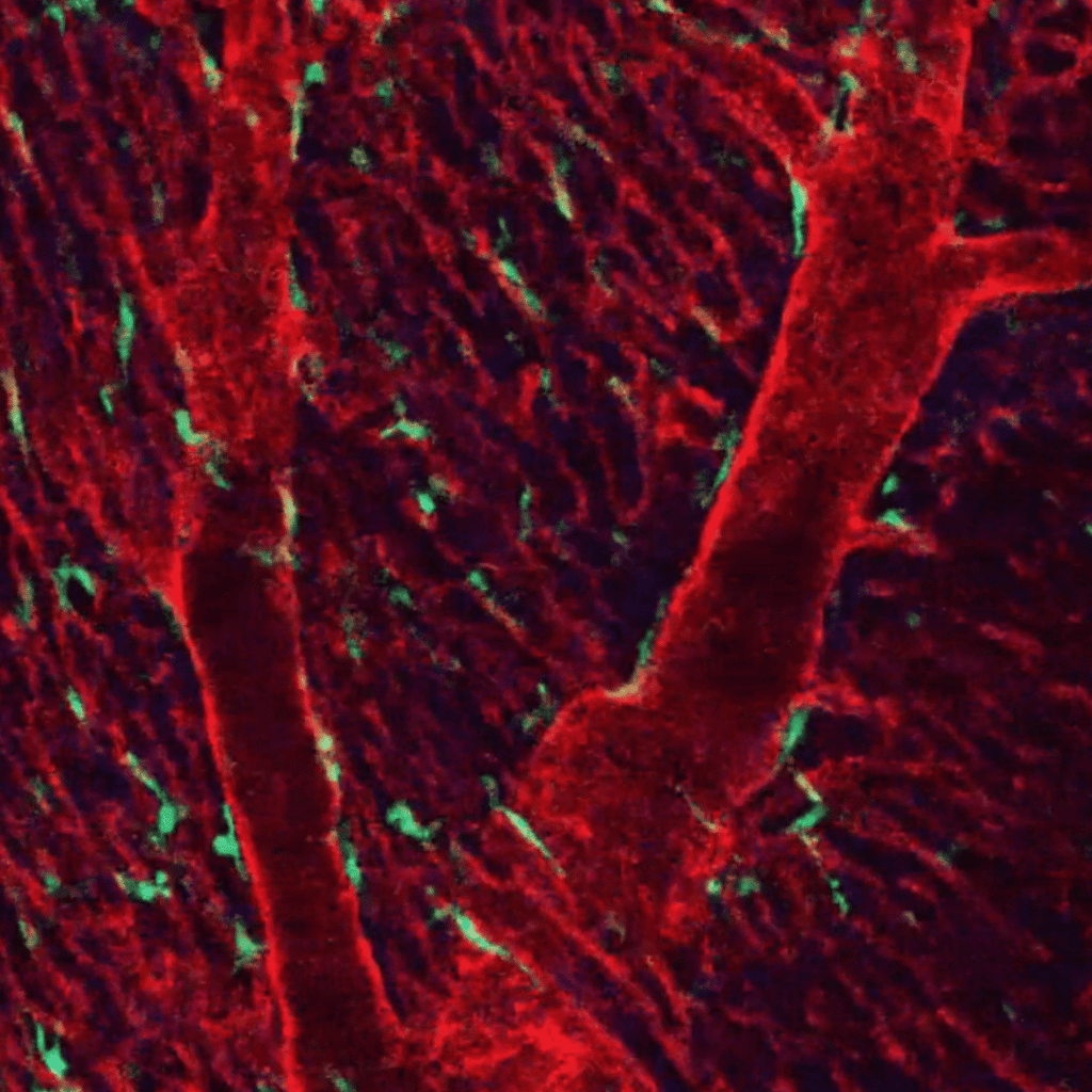
This study introduces a better intravital heart imaging protocol that uses a suction-based cardiac window and AI-driven motion compensation to stabilize the beating mouse heart. Researchers used confocal and two-photon microscopy in live LysM-eGFP mice to clearly see immune cell movement, RBC trafficking, and cardiac muscle structures in real time. The method greatly reduces motion artifacts, allowing for precise measurement of cell dynamics and detailed imaging of sarcomeres and fibrillar organization.
Interested in Intravital Microscopy?
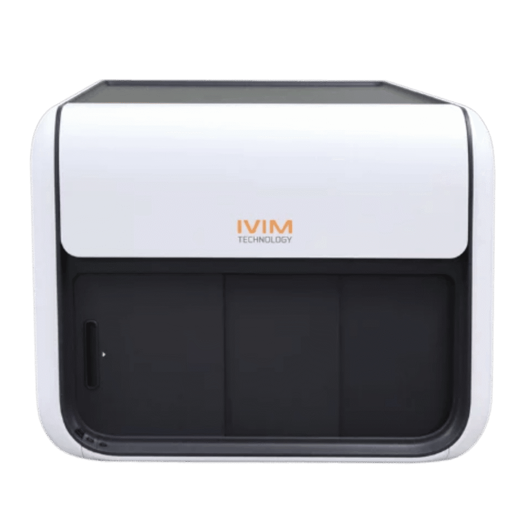
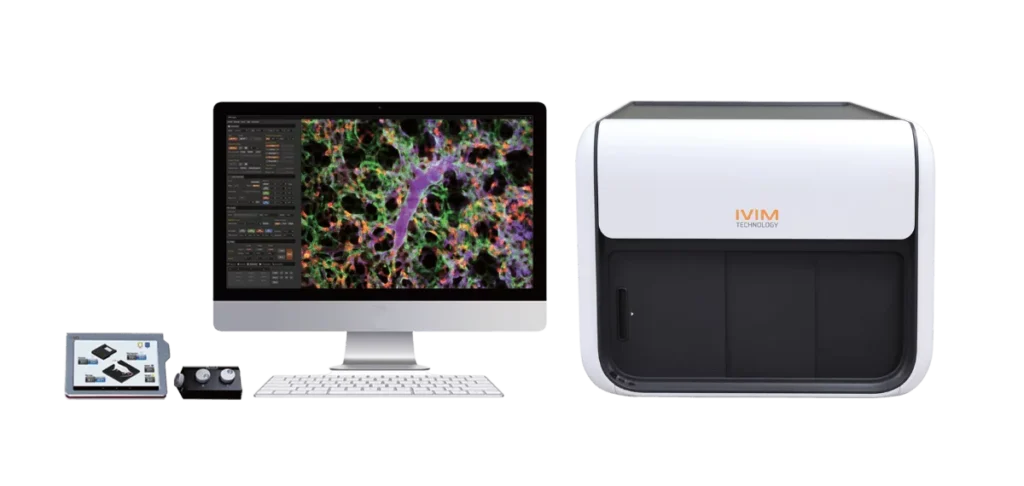
Check out the IVIM System
*Information presented on this page and website reflects the data, product specifications, application information, and regulatory status available at the time of publication. Product features, specifications, availability, pricing, and regulatory information may change without notice. Please consult the manufacturer or contact Scintica for the most current information.
*This content is provided for informational purposes only and is based on information available at the time of publication. While reasonable efforts have been made to ensure accuracy, product specifications, performance data, regulatory status, availability, and other details may change without notice. Scintica makes no representations or warranties regarding the ongoing accuracy or completeness of the information presented and recommends verifying all information with the manufacturer before making purchasing or research decisions.

Enter your details and we’ll email the requested brochure to you.