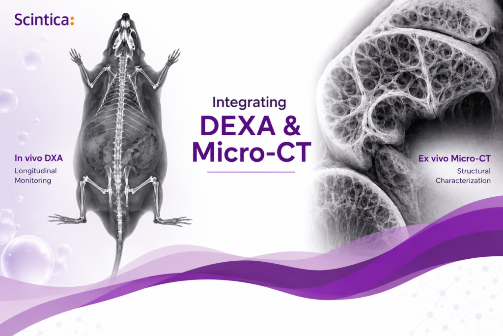
Product Highlight
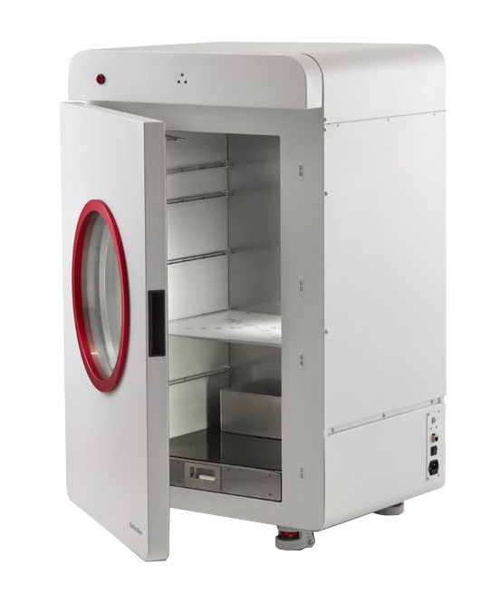
iNSiGHT DXA
The iNSiGHT DXA system is designed for preclinical bone mineral density and body composition assessment.
View key features
- The fastest in vivo scan time (<25 seconds)
- Low-dose ionizing radiation
- Quantification of BMD, BMC, fat mass, and lean mass
- Whole-body and regional analysis
- Large field of view (16.5 x 25.5 cm)
- Animal welfare-focused workflow design
- Preclinically validated against gold standard methods for BMC and body composition (chemical carcass analysis and bone ashing)
View workflow advantages
The iNSiGHT DXA supports efficient longitudinal studies by enabling repeated measurements with minimal animal burden. Additional workflow advantages include:
- Self-shielded cabinet design
- Active gas scavenging for anesthesia workflows
- High throughput acquisition
- User-friendly software
Product Highlight
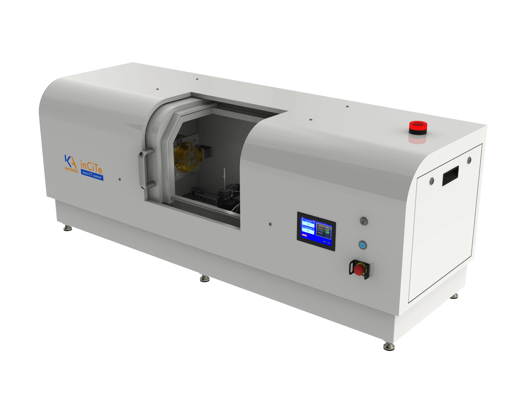
inCiTe™ 3D Micro-CT
The inCiTe™ 3D X-ray Microscope is designed for high-resolution ex vivo characterization of tissue microarchitecture and structural organization.
View key capabilities
- Phase contrast imaging for better edge enhancement
- Submicron resolution at highest magnification
- Advanced imaging of implants and biomaterials interfaces
- 3D volumetric imaging
- Simplified workflow
