Submitted by Dr. Gracie Vargas, Professor, Neuroscience Cell Biology & Anatomy and Director of the Advanced Bio-Optics Imaging Laboratory, University of Texas Medical Branch, Galveston, TX.
The biomedical engineering and women’s research teams headed by Drs. Massoud Motamedi, Dr. Gracie Vargas, and Dr. Kathleen Vincent at the University of Texas Medical Branch Center have a collective interest in image-based assessment of vaginal and rectal epithelial injury and inflammation. The occurrence of injury or inflammation is associated with potential compromise of the barrier function of the vaginal and rectal mucosal surfaces. Defects in barrier function or recruitment of target cells which may occur with inflammation or healing of injuries are of concern as they could increase risks for transmission and infection by HIV and other pathogens. Furthermore, there is a paucity of methods that can provide indication of vaginal and rectal epithelial injury and inflammation in vivo or in real time, and, in particular, to detect and monitor mucosal health in response to topical or systemic drugs aimed for prevention of infection. This remains true not only for monitoring of effects in humans but also as tools in the development of preventive approaches in small and large animal models.
For the last several years, this collective team of biomedical engineers and women’s studies researchers have led efforts to 1) understand the mucosal changes associated with the use of topical, systemic, or device-delivery of HIV prevention agents and 2) identify markers, including image-based markers of inflammation and injury to these surfaces. Image tools that have been central to our efforts have included the real-time optical imaging tools of confocal endomicroscopy [1-3] and optical coherence tomography [4-5].
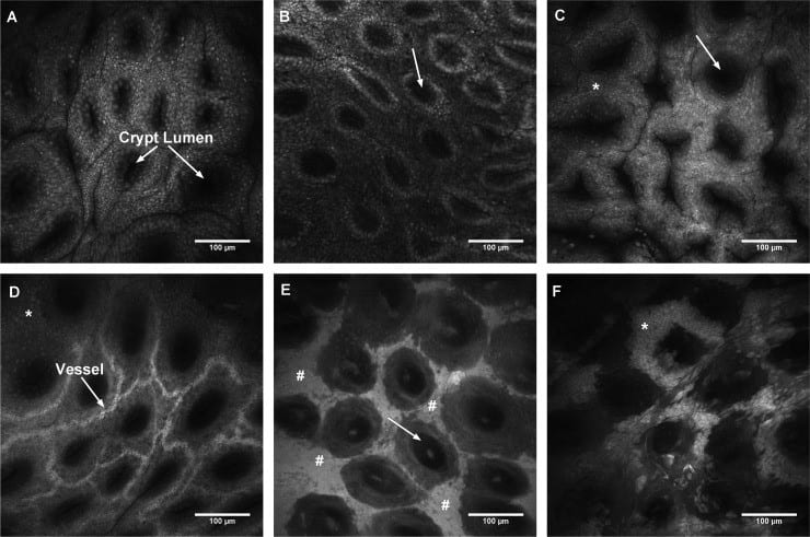
Confocal endomicroscopy used in this research has included use of the OptiScan FIVE.1 confocal endomicroscope [1,2] and most currently, the ViewnVivo (FIVE.2) endomicroscope. These in vivo microscopes have allowed for rapid and longitudinal imaging of the vaginal and rectal mucosa in a large animal model used in our studies. In vivo microscopy has allowed identification of surface structural and functional features that indicate injury or compromise (Figure 1). Furthermore, with appropriate staining, inflammatory infiltrates may be identified and potentially used to monitor presence of inflammation and impact on mucosal health. Our group anticipates this tool to continue to be a valuable component of our studies in women’s health and HIV prevention.
Figure 1: Features of the ovine rectal mucosa identified prior to and following epithelial disruption from topical agents. Panels A-D show structural features made possible from a topical dye (Acriflavine) while E-F show compromise of the barrier function through leakage of a paracellular dye (NaFl).
{From: Vargas G, Vincent KL, Zhu Y, Szafron D, Brown TC, Villareal PP, Bourne N, Milligan G, Motamedi M, In vivo rectal mucosal barrier function imaging in a large animal model using confocal endomicroscopy: implications for injury assessment and use in HIV prevention studies, Antimicrobial Agents and Chemotherapy, 2016 May 16. pii: AAC.00134-16.}
Professor
University of Texas Medical Branch
 Dr. Gracie Vargas, Professor, Neuroscience Cell Biology & Anatomy, is a biomedical engineer with expertise in bio-optical imaging and biophotonics. She is Director of the Advanced Bio-Optics Imaging Laboratory at UTMB, focusing on the integration of engineering principles and advanced imaging with the biomedical sciences. Her laboratory conducts research applying the techniques of nonlinear optical microscopy and confocal endomicroscopy for the study of injury and disease processes and as potential tools in noninvasive diagnostics. Major areas of current focus are in epithelial cancer diagnostics, endomicroscopic evaluation of epithelial injury for assessing potential toxicity of candidate HIV-prevention agents, and large-scale microscopy enabled by tissue optical clearing. She has had funding awards from sources such as the National Institutes of Health, National Science Foundation, and Cancer Prevention Research Institute of Texas. She is also an active educator, mentoring trainees at all levels from high school graduate and medical school level, and has a specific interest is in advancing the representation from historically underrepresented groups in science and engineering. Dr. Vargas is also highly active in scholarly service at the national level serving as a standing member for NIH study sections among other activities.
Professor
University of Texas Medical Branch
 Dr. Massoud Motamedi is a Professor and the Vice Chair for Research in the Department of Ophthalmology and Visual Sciences at the University of Texas Medical Branch (UTMB) in Galveston, TX. Dr. Motamedi received his undergraduate and graduate training in electrical engineering with concentration in biomedical engineering in the Department of Electrical Engineering at the University of Texas at Austin. He is a recognized expert in the fields of biophotonics, imaging and biomedical engineering. He has made significant contributions to the development and applications of novel imaging and sensing platforms for biomedical diagnosis and sensing. At UTMB, he has successfully directed research programs with a focus on biomedical imaging, sensing and applications of photonics and imaging in biology and medicine. As part of these efforts he has interdisciplinary research projects and training programs for graduate and medical students, residents and fellows at UTMB while contributing to the development of several imaging techniques and their applications in the field of medicine. Dr. Motamedi’s research effort has been supported by various funding agencies including NIH, NSF, DOD and private foundations. He is a fellow in the American Institute for Medical and Biological Engineering College of Fellows.
Associate Professor
University of Texas Medical Branch
 Kathleen Vincent, MD., Associate Professor of Obstetrics and Gynecology, is a board certified and practicing obstetrician/gynecologist with a degree in Chemical Engineering. Dr. Vincent has conducted biomedical research for over 20 years and currently maintains a research program in women’s health, centered on the use of biomedical engineering technologies applied to problems in women’s health. Her research focuses on the use of advanced imaging and biomedical engineering to improve the health of women, in particular, thfemale reproductive health. She has led efforts to explore the use of emerging endoscopic imaging techniques for the evaluation of injury and disease, and in the last 13 years evaluating drug toxicity and the integrity of vaginal and rectal epithelial barrier in both small and large animal models. During this time, she has worked with industry toward the development of safe and effective topical microbicides aimed at HIV prevention. Dr. Vincent has also translated advanced imaging techniques into clinical research and is funded to conduct clinical studies using optical coherence tomography and other methods to study the use of intravaginal rings to prevent HIV and for medical treatment of urinary incontinence. This research has been largely funded by NIH.
References:
1.Vargas G, Patrikeev I, Wei J, Bell B, Vincent K, Bourne N, Motamedi M, Quantitative assessment of microbicide-induced injury in the ovine vaginal epithelium using confocal microendoscopy, BMC Infectious Diseases 2012, 12:48 (29 February 2012) PMID: 2237579
2.Vargas G, Vincent KL, Zhu Y, Szafron D, Brown TC, Villareal PP, Bourne N, Milligan G, Motamedi M, In vivo rectal mucosal barrier function imaging in a large animal model using confocal endomicroscopy: implications for injury assessment and use in HIV prevention studies, Antimicrobial Agents and Chemotherapy, 2016 May 16. pii: AAC.00134-16.
3.Vargas G, Vincent KL, Wei J, Bourne N, Motamedi M, Topical injury evaluation of the murine colorectal mucosa using confocal endomicroscopy: a valuable method for assessing mucosal injuries associated with risk of pathogen transmission, J Microsc. 2016 Jun 28. doi: 10.1111/jmi.12438. [Epub ahead of print] PMID: 27351717
4.Vincent, K.L., G. Vargas, J. Wei, N. Bourne and M. Motamedi, Monitoring vaginal epithelial thickness changes noninvasively in sheep using optical coherence tomography. American journal of obstetrics and gynecology, 2013 Apr;208(4):282.
5.Vincent, K.L., G. Vargas, N. Bourne, V. Galvan-Turner, J. I. Saada, G. H. Lee, E. Sbrana and M. Motamedi, Image-Based Noninvasive Evaluation of Colorectal Mucosal Injury in Sheep After Topical Application of Microbicides. Sexually transmitted diseases, 2013. 40(11): p. 854-859.