Application Note: Bioprinting 3D Multi-cellular Cartilage Spheroids Using Laser-Assisted Technology
References
Hall GN, Fan Y, Viellerobe B, Iazzolino A, Dimopoulos A, Poiron C, Clapies A, Luyten FP, Guillemot F, Papantoniou I. Laser-assisted bioprinting of targeted cartilaginous spheroids for high density bottom-up tissue engineering. Biofabrication. 2024 Aug 22;16(4). doi: 10.1088/1758-5090/ad6e1a. PMID: 39136309.
Multicellular Spheroids
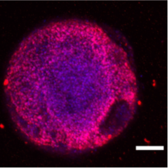
Figure 1– Embryoid Body (EB) Oct4 DAPI. Scale bar 1 mm.
Organoids are three-dimensional tissue cultures that replicate the structure and function of organs and created from stem cells such as embryonic stem cells, adult stem cells, or induced pluripotent stem cells (iPSCs). They are grown in a supportive matrix and can also be developed by coculturing epithelial progenitors with mesenchymal and endothelial cells. Organoids’ 3D microenvironment allows for both cell-cell and cell-matrix interactions that heavily mimic the native tissue. Because of this, they have shown a lot of potential in the fields of drug discovery, personalized diagnostics, cell therapy, and modeling human-specific aspects of development and disease1.
Cartilaginous Spheroids
Cartilaginous spheroids are multicellular 3-dimensional constructs that can be used for tissue engineering, cartilage regeneration and as in vitro models for a variety of studies. Unlike other spheroids, cartilaginous microtissues tend to be much larger structures. Producing spheroids have largely relied on random self-assembly of within constrained containers, with limited control over size, and architectural features. Increase precision of cartilaginous spheroid assembly with specific architectural features and sizes would require a more automated and controlled biofabrication methods, which became possible with laser-assisted bioprinting.
Advantages of Laser-Assisted Bioprinting
Previous bioprinting technologies have been used for producing multicellular spheroids, but they come with several drawbacks. Extrusion-based bioprinting, which encapsulates spheroids in hydrogel bioink, often produce low spheroid densities and limited control over spheroid localization within the final construct. In addition, this technology has the added issue of nozzle clogging, particularly when spheroids fuse into larger agglomerates prior to bioprinting. Although aspiration-based techniques have been developed for picking up and depositing single spheroids to combat this issue, this bioprinting method is still limited in throughput.
Laser-assisted bioprinting (LAB), on the other hand, is a nozzle-free technique and offers greater precision in manipulating cell suspensions at a single-cell resolution. This nozzle-free process is of interest for spheroid bioprinting to avoid the issues seen with extrusion-based methods. LAB works by focusing a pulsed laser on a donor substrate coated with a metallic layer and bioink. The laser energy causes the metal to ablate, creating a plasma that generates a cavitation bubble. This bubble collapses, producing a liquid jet that deposits bioink droplets containing a defined number of cells onto a receiver, enabling precise spheroid deposition and helps to overcome the limitations of traditional methods.
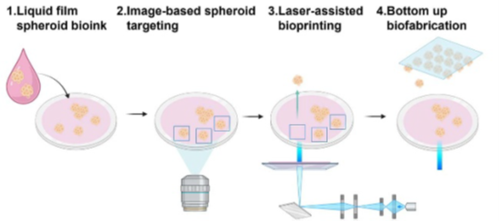
Figure 2– Spheroid laser-assisted bioprinting: Cell spheroids are imaged, targeted, and transferred by the laser to form a predesigned pattern.1
Proof of Concept
Hall et al. demonstrated the successful bioprinting of cartilaginous spheroids formed from human periosteum-derived cells (hPDCs). The spheroids maintained high cell viability and retained their capacity for chondrogenic differentiation post-printing (Fig. 3). Smaller spheroids (100–150 μm in diameter) were successfully bioprinted using the Laser-Induced Forward Transfer (LIFT) method. However, printing larger spheroids proved challenging. A novel method was implemented, called Laser-Induced Propulsion of Mesoscopic Objects (LIPMO), enabling bioprinting of larger spheroids (up to 300 μm). The bioprinting process, combined with computer-aided image analysis allowed for successful fabrication of high-density, multi-layered spheroid populations.

Figure 3– Cartilaginous spheroids was confirmed to be viable after printing with immunostaining and were able to undergo differentiation and maturation. Reproduced from Hall et.al. (2024).
Next Generation Bioprinting System (NGB-R™) by Poietis™

Figure 4– NGB-RTM Robotic-Assisted LAB System.
The LAB system used in this study is Next Generation Bioprinting systems (NGB-R), designed and manufactured by Poietis™. NGB-RTM is a multimodal, 3D bioprinting platform designed and developed to print live tissues and organs. The system combines laser-assisted, and extrusion bioprinting, enabling researchers the versatility of bioprinting (from single cells to 3D spheroids) and offers the possibility of using a large number of biomaterials and hydrogels. Integrated robotics and microscopes enables automating the entire bioprinting process with high resolution and speed.