Utilizing Ultrasound Imaging & Shear Wave Elastography to Assess Blood Arteriovenous Fistulas & Abdominal Aortic Aneurysms
Ultrasound imaging combined with shear wave elastography (SWE) is an advanced technique increasingly used in clinical and preclinical research for assessing blood vessel pathologies and advanced interventions, including arteriovenous fistulas (AVFs) and abdominal aortic aneurysms (AAAs). This method integrates the high-resolution imaging capabilities of traditional ultrasound with the biomechanical assessment provided by SWE, offering a comprehensive tool for advanced, non-invasive blood vessel evaluation.
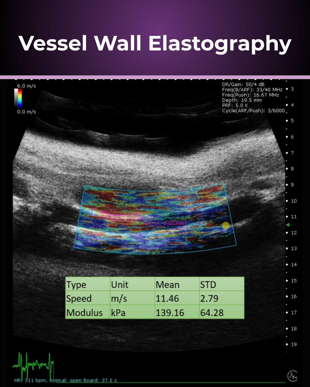
Ultrasound imaging is widely utilized in both clinical and preclinical settings due to its ability to provide real-time, high-resolution images of soft tissues. It uses high-frequency sound waves that penetrate the body, reflect off tissues, and return to the transducer, creating detailed images based on the differing acoustic properties of tissues. In vascular research, ultrasound imaging allows for the visualization of vascular wall thickness, diameter, and blood flow. Applications for standard ultrasound imaging include confirming fistula presence, measuring venous and arterial blood flow rates, or confirming the diameter or volume of a developing aneurysm. Microvascular perfusion can also be measured when using a contrasting agent such as microbubbles. SWE is a functional imaging technique that measures the stiffness or elasticity of tissues. It works by generating mechanical shear waves within the tissue using acoustic radiation forces. These waves propagate through the tissue, and their speed is directly related to the tissue’s stiffness. The ultrasound transducer detects these waves and calculates the elasticity modulus, producing a quantitative map (elastogram) of tissue stiffness. In the context of blood vessels, tissue stiffness is a crucial biomarker that could be indicative of many pathologies, including elevated total peripheral resistance. Therefore, SWE can provide valuable insights into the biomechanical properties of blood vessels.
Applications in Preclinical Vascular Research
In preclinical vascular research, combining ultrasound imaging with SWE offers several advantages:
1. Vessel Detection and Characterization.
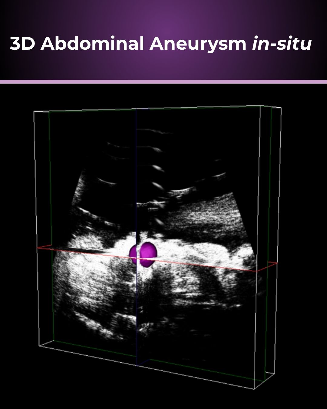
- Structural Imaging: Ultrasound provides detailed images of the vessel’s structure, allowing researchers to detect and monitor vessel aneurysm diameters in animal models.
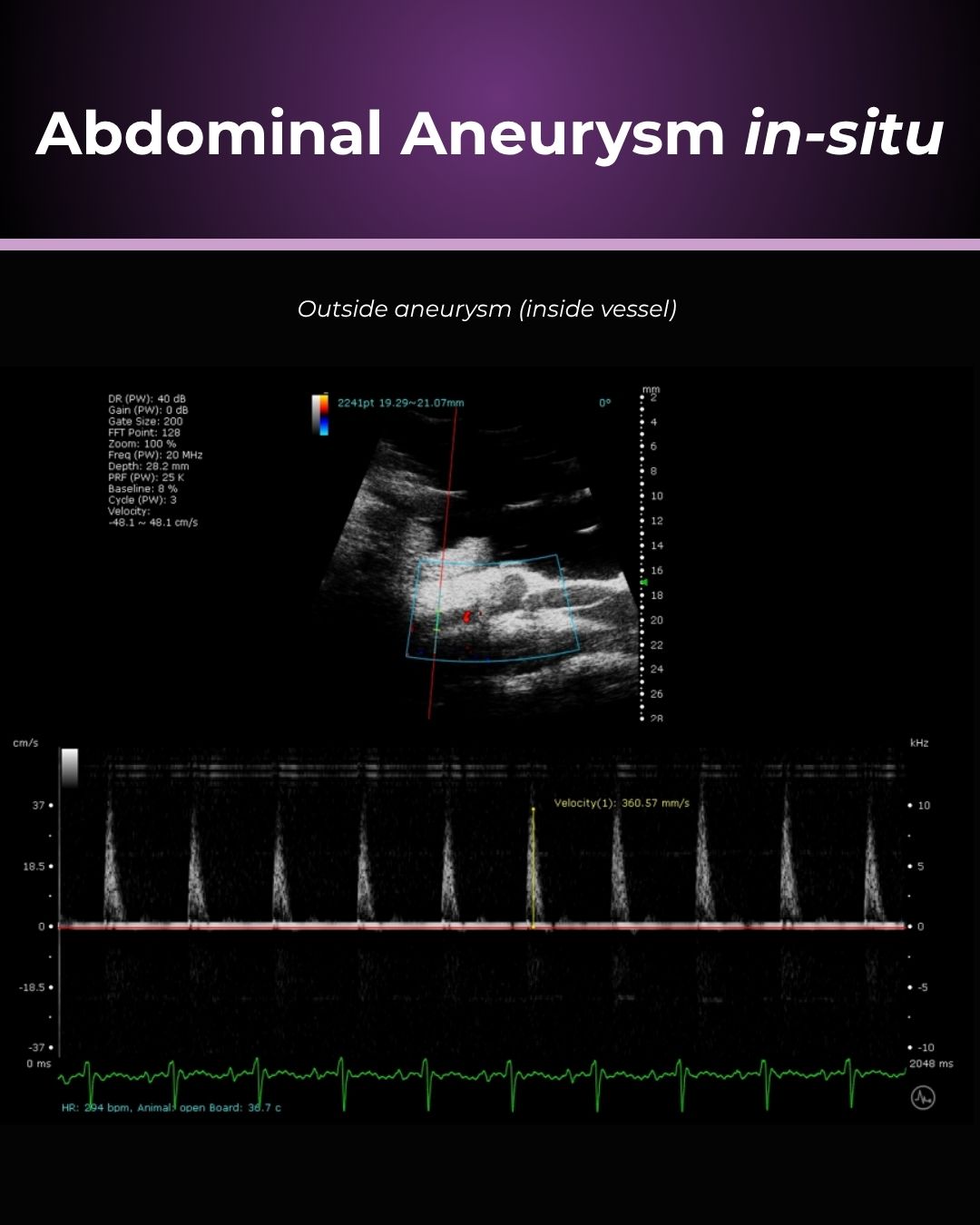
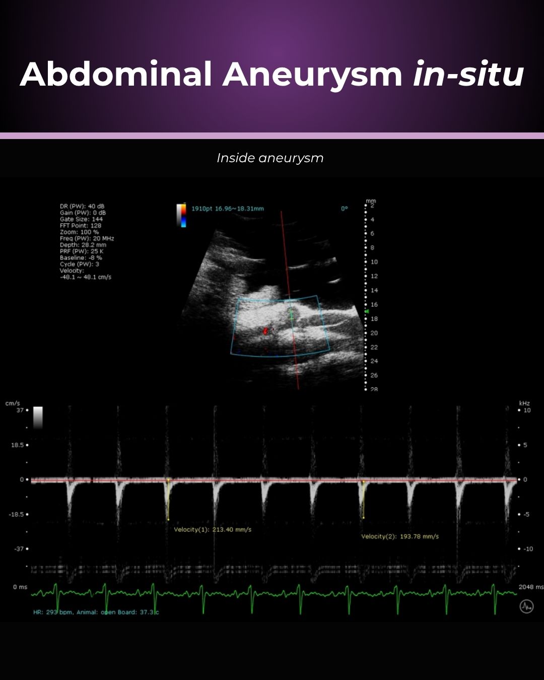
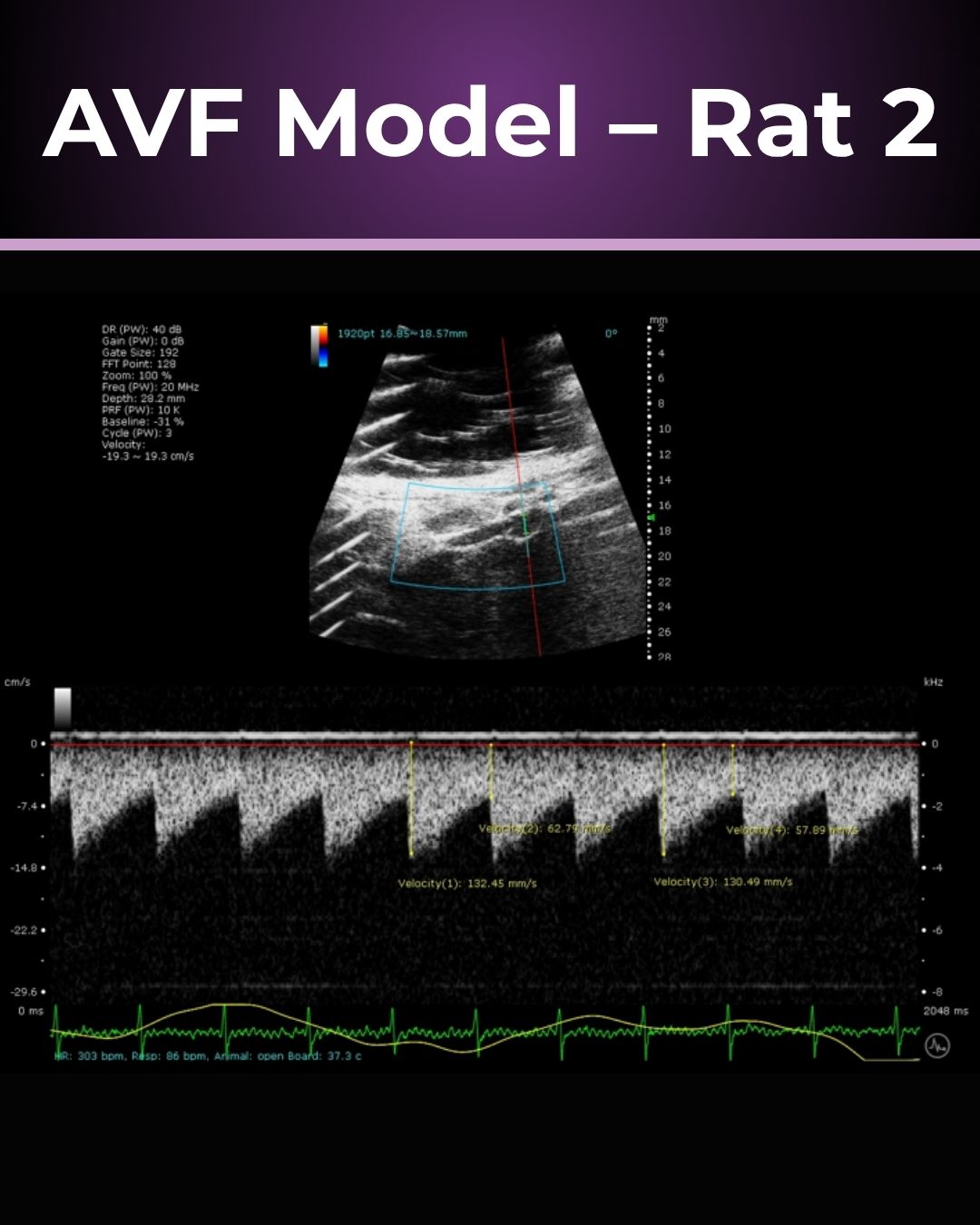
- Blood Flow Measures: Blood flow measurements by colour Doppler provide flow direction and amplitude data that can provide crucial information about aneurysm thrombosis risk, fistula efficacy, and maturation.
- Elasticity Mapping: SWE adds a layer of functional information by mapping tissue stiffness. This is particularly useful for understanding vessel wall stiffness that can otherwise be difficult to measure non-invasively.
2. Monitoring Vessel Characteristics and Treatment Response Evaluation
- Longitudinal Structural Imaging: The non-invasive nature of ultrasound and SWE allows for longitudinal measures, improving your statistical power while also reducing animal recruitment.
- Therapeutic Efficacy: Ultrasound and SWE can be used to assess the effectiveness of your intervention, whether it’s the effect of a drug on vessel diameter response or wall stiffening interventions.
Advantages of Using Ultrasound with Shear Wave Elastography in Preclinical Research
Non-Invasive
Both ultrasound imaging and SWE are non-invasive, reducing the requirement for biopsies and minimizing harm to animal subjects.
Real-Time Monitoring
These techniques provide real-time data, allowing for immediate assessment and decision-making in experimental studies.
Quantitative Data
 SWE provides quantitative measurements of tissue stiffness, which can be objectively analyzed and compared across studies.
Cost-Effectiveness
Ultrasound and SWE are relatively cost-effective compared to other imaging modalities like MRI or CT, making them accessible for routine use in pre-clinical research.
Product Highlight:
Prospect T2
Preclinical Ultrasound System
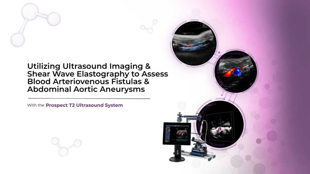
The Prospect T2 is an innovative high-frequency ultrasound system designed specifically for in vivo preclinical imaging in small animals such as mice and rats. This compact and cost-effective tablet-based system provides high-resolution images (up to 30 µm) and advanced capabilities to monitor changes in hemodynamics and observe anatomical structures in real-time.
View key capabilities
- Compact & Small Footprint
- Add-on Hardware/Software Options
- Multiple Probes & Platforms
- Simplified Image Guided Injection Mount