Title:Â Redefining Cardiovascular Discovery Through Multimodal Imaging
Date: April 23–26, 2026
Location: American Physiology Summit (APS) in Minneapolis
Presenter: Dr. Sadi LoaiÂ
Recorded at the 2026 American Physiology Summit (APS) in Minneapolis, this presentation by Sadi Loai, Ph.D., Senior Product Manager at Scintica, explores how multimodal imaging can provide a more comprehensive view of cardiovascular structure, function, perfusion, metabolism, and disease progression in preclinical research.
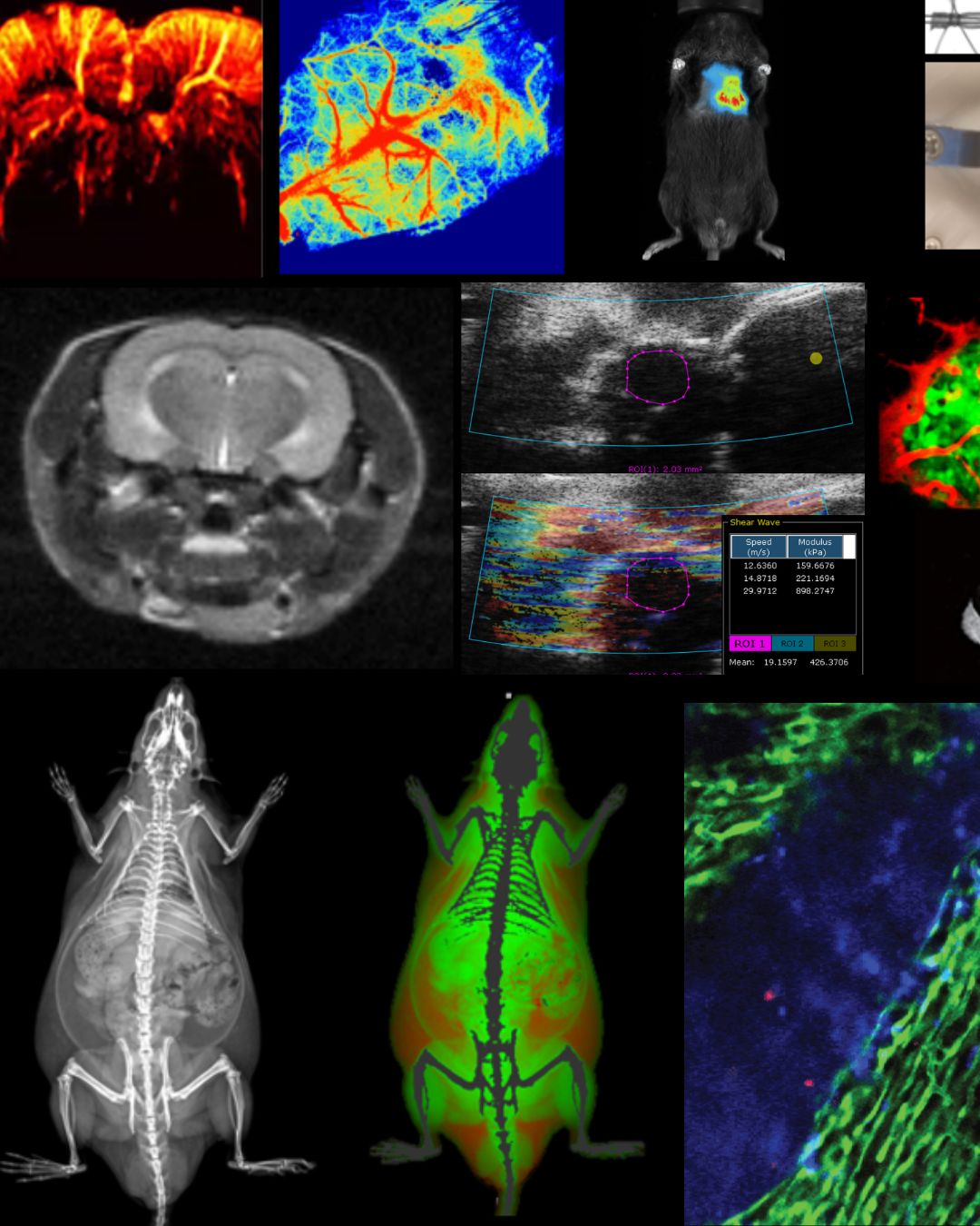
Preclinical cardiovascular research is undergoing a rapid transformation as advanced imaging technologies converge to deliver deeper, more dynamic insights into heart and vascular biology. This workshop explored how modalities such as high-frequency ultrasound, MRI, DEXA, optical imaging, laser Doppler, and emerging techniques are reshaping study design, data integration, and translational impact.
Attendees learned how multimodal approaches enable comprehensive assessment of cardiac structure, function, perfusion, metabolism, and disease progression in vivo. Through real-world examples and practical guidance, the session highlighted how combining technologies can improve data quality, reduce animal numbers, and accelerate cardiovascular discovery from mechanism to therapy.
What You Will Learn:
- Why multimodal imaging matters in diabetic heart disease
- Complete mouse cardiac function analysis with ultrasound
- MRI and DXA for vascular and metabolic insight
- Real-time cellular imaging of the beating heart
Preclinical Imaging Portfolio
The goal of multimodal imaging is to acquire as much data as possible with respect to a biological question, to allow for a more holistic examination then would be possible with a single imaging modality, as well as to approach the biological question from multiple scale levels.