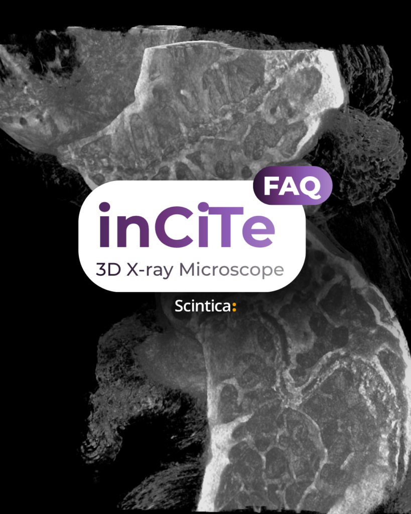
FAQ: inCiTe™ 3D X-ray Microscope
The inCiTe™ 3D X-ray Microscope combines high-resolution micro-CT with propagation-based phase contrast to help researchers visualize low-density biological structures and biomaterials that may be difficult to distinguish using absorption imaging alone. Explore the questions below to determine whether inCiTe™ 3D could support your research.
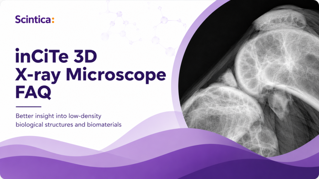
Frequently Asked Questions
What makes the inCiTe™ 3D different from a conventional micro-CT system?
Conventional micro-CT primarily creates contrast from differences in X-ray absorption. This works especially well for highly attenuating materials such as mineralized bone, but low-density structures with similar absorption properties can be difficult to resolve/differentiate. inCiTe™ 3D adds propagation-based phase contrast, which is sensitive to changes in how X-rays are refracted as they pass through a sample. This can improve the visibility of boundaries, interfaces, and subtle structural variations in weakly absorbing materials such as soft biological tissues, polymers, and tissue-engineered scaffolds.
The inCiTe™ 3D system uses KA Imaging’s direct-conversion BrillianSe™ detector to achieve this sought-after capability and high level of performance.
What types of biomedical samples are best suited for inCiTe™ 3D?
The strongest candidates are ex vivo samples containing low-density structures, internal interfaces or materials that are difficult to distinguish using absorption contrast alone. Potential applications include:
Articular cartilage, meniscus and osteochondral specimens
Tendon, ligament and bone-soft tissue interfaces
Tissue-engineered scaffolds
Polymer-tissue and implant-tissue interfaces
Porous biomaterials and regenerative-medicine specimens
Samples undergoing structural deformation or internal damage
Suitability ultimately depends on the sample dimensions, composition, preparation, and the feature the researcher needs to visualize.
Can inCiTe™ 3D image bone and soft tissue within the same specimen?
Yes. inCiTe™ can image both mineralized and lower-density structures within the same specimen. This makes it particularly interesting for research involving osteochondral tissue, joint structures, fracture repair, bone-implant interfaces where bone is only one part of the biological question. However, phase contrast does not automatically make every soft tissue equally visible. Image quality will depend on the size of the structure, its density relative to neighbouring materials, sample preparation and scan conditions. A feasibility scan is recommended when the target structure has very low inherent contrast.

Who would benefit most from the inCiTe™ 3D X-ray Microscope?
inCiTe™ 3D is particularly relevant for researchers examining specimens that contain both mineralized and lower-density tissues:
Musculoskeletal researchers
evaluating the relationships between bone, cartilage, tendons and ligaments during healing or disease.
Biomechanics researchers
investigating bone, muscle and connective tissue under relevant loads.
Cancer researchers
assessing tumour structure and blood vessel architecture when mineralized tissue is nearby.
Regenerative medicine and biomaterials researchers
studying how engineered materials interface and integrate with adjacent tissues over time.
When conventional imaging cannot adequately visualize both tissue types in one scan, researchers may use contrast agents, tissue staining, histology or additional imaging methods. These approaches provide valuable information but require additional preparation, add complexity to the workflow, and may reduce the ability to image the same specimen repeatedly.
For researchers seeking comprehensive, non-destructive evaluation, inCiTe™ 3D offers an opportunity to obtain more structural information during a single imaging session while preserving the specimen for subsequent analysis.
Does inCiTe™ 3D replace a conventional bone micro-CT system?
Not in every application. Conventional systems with established bone-analysis workflows may remain the preferred choice for laboratories focused primarily on routine bone morphometry or high-volume scanning.
inCiTe™ 3D is most differentiated when the research question extends beyond mineralized bone to include cartilage, weakly absorbing tissues, tissue-material boundaries, low-density biomaterials, scaffold architecture or internal interface failure.
Rather than asking, “Do I need micro-CT?”, ask, “Which structures and interfaces do I need to distinguish?”. The answer to the latter will help guide your product research, grant applications, and purchasing decisions.
Do samples need to be stained or treated with a contrast agent?
Not necessarily.
Phase contrast may improve the visibility of naturally occurring boundaries without requiring a traditional X-ray contrast agent. This can help preserve the specimen for subsequent analysis and reduce the preparation burden for certain applications.
Staining or another preparation method may still be beneficial when the target tissue is highly homogeneous, surrounded by fluid with a similar density, very small, or when greater segmentation contrast is required for quantitative analysis. Our team can help assess whether native phase contrast, optimized preparation, or contrast enhancement is most appropriate for a particular sample.
What information can researchers obtain from an inCiTe™ dataset?
inCiTe™ produces high-resolution three-dimensional datasets that can be reconstructed, visualized and analyzed using compatible software workflows. Depending on the specimen and image quality, researchers may be able to evaluate:
- Internal morphology and three-dimensional architecture
- Material or tissue thickness
- Pore size, porosity and pore interconnectivity
- Branching geometry, scaffold continuity
- Internal voids, cracks and defects
- Tissue-material contact and interface integrity
- Dimensional changes with mechanical loading
The exact quantitative endpoints should be established during study planning. Not every visible boundary will necessarily support reliable automated segmentation, so analysis feasibility should be evaluated alongside imaging feasibility.
Is inCiTe™ 3D intended for in vivo animal imaging?
Static, ex vivo specimens
inCiTe™ 3D is best positioned for static, ex vivo biological specimens, biomaterials and engineered constructs. It is not designed as a routine longitudinal live-animal micro-CT platform.
Its ex vivo workflow is valuable for endpoint studies because the specimen can be examined non-destructively in three dimensions before proceeding to histology, mechanical testing, or other destructive analyses. Imaging may also help researchers select the most informative regions for subsequent sectioning.
How do I determine whether my sample is a good fit?
The most reliable approach is to begin with a sample-feasibility assessment. Our team will review:
- The sample type, composition and dimensions
- The structure or interface you need to visualize
- Your current imaging method and its limitations
- Sample fixation, hydration or embedding conditions
- The approximate size of the feature of interest
- Whether quantitative segmentation is required
- Any need to preserve the specimen for downstream testing
Representative imaging can then be used to determine whether inCiTe™ provides meaningful additional information before you commit to a larger study or system evaluation.
Related Resources
Explore related product and application resources:
Can conventional micro-CT answer your entire research question?
Tell us what you are trying to visualize. Our imaging team can review your specimen, recommend an initial workflow, and determine whether phase-contrast imaging could reveal additional structural information.

Titanium implant into a bone construct