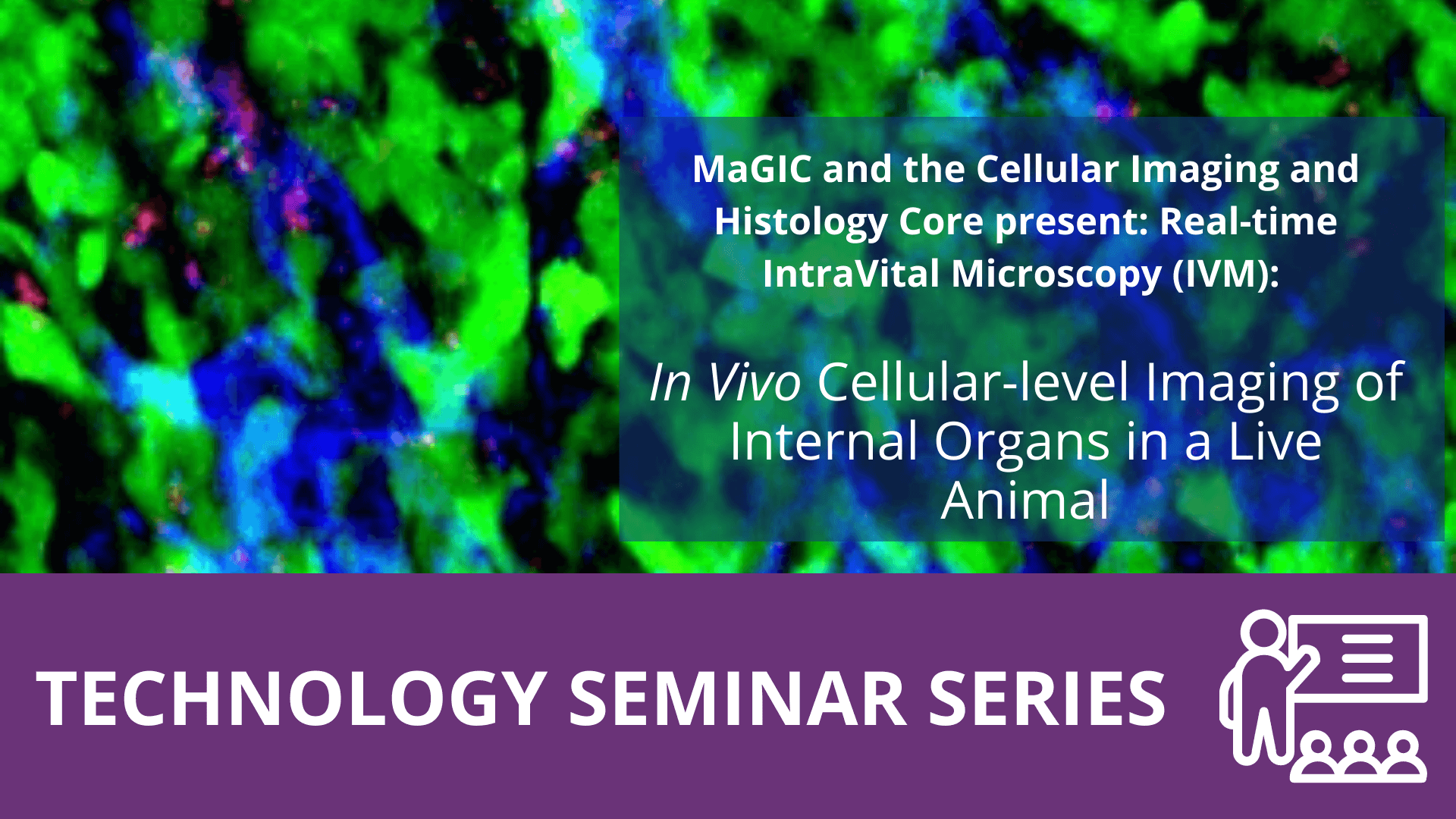
Intravital microscopy is a unique imaging technique to visualize various in vivo cellular-level dynamics such as cell trafficking, cell-to-cell or cell-to-microenvironment interactions in a live animal. Intravital imaging of cellular dynamics in a natural physiological microenvironment can provide unprecedented insights in the dynamic pathophysiology of human diseases those were impossible to obtain through conventional histological observation of ex vivo sample or in vitro culture system. During the last decade, the intravital microscopy has become a highly valuable, indispensable technique in wide areas of biomedical sciences such as immunology, neuroscience, developmental and tumor biology. Notably, in vivo visualizations of gene expression, protein activity, cell trafficking, cell-cell / cell-microenvironment interactions and various physiological responses to external stimuli have been successfully achieved. Additionally, it’s a unique tool for the development of new therapeutics and diagnostics by providing improved accuracy and reliability in in vivo target validation with delivery monitoring and efficacy assessment. It has been used to directly analyze the delivery and efficacy of new biopharmaceuticals such as antibodies, cell therapy, gene therapy, nucleic acids and exosome in an in vivo microenvironment.
In this talk, IVIM Technology’s All-in-One real-time intravital two-photon and confocal microscopy system will be introduced. The imaging system has been extensively optimized for in vivo cellular-level imaging of internal organs in live animal model for various human diseases. It can acquire a real-time multi-color sub-micron resolution microscopic images in a live animal model with automatic motion compensation, enabling direct imaging analysis of complex cancer immune-microenvironment consisted of various immune cells, stromal cells, vascular cells and extracellular matrix.