Tomographic imaging is a cornerstone of advanced medical imaging. It has transformed our ability to visualize the internal structures of organisms. This has led to a new diagnostic era. Clinicians can now readily diagnose various conditions non-invasively, from bone fractures to cancer. But the relevance of tomography isn’t exclusive to the clinic. Researchers also make extensive use of tomographic imaging modalities in preclinical settings.
The Working Principles of Tomographic Imaging
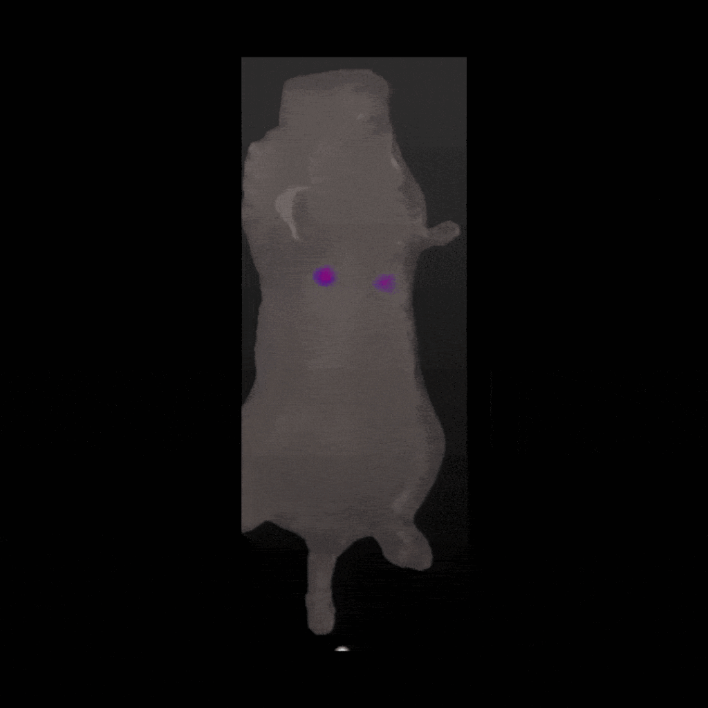
Tomographic imaging is a sophisticated technique that goes beyond simply capturing data. It involves capturing data from multiple perspectives, which allows for a more comprehensive and accurate image to be created. This is achieved through the use of mathematical algorithms that piece together the data collected from different angles, resulting in a detailed representation of internal structures. Unlike traditional optical imaging systems that use straight lines or ionizing radiation, tomographic imaging uses diffusive optical strategies. This means it uses light to examine the inside of objects or organisms. Using light offers a non-invasive and safe way to get detailed images. As it propagates through the sample, the light is scattered off internal structures. It’s this scattering effect that provides such clear data about what’s inside the object under test. Algorithmic processing of this data enables precise reconstructions of the internal environment. A further benefit of tomographic imaging is its ability to provide real-time images. This is especially useful in medical settings, where it can help track diseases or see how treatments are working.Comparison with Other Techniques
Traditional imaging methods (i.e. X-ray imaging) primarily offer a two-dimensional view and rely on ionizing radiation. In contrast, tomographic imaging, especially techniques like diffuse optical tomography (DOT) and optical coherence tomography, provides a three-dimensional, high-resolution insight without the risks associated with radiation. This depth is crucial when studying intricate biological systems or soft tissue. In preclinical research, the superiority of tomographic imaging becomes evident. Systems like the Newton 7.0, an optical bioluminescence and 3D tomographic imaging system, are designed for such specialized tasks.Applications in Preclinical Settings
The applications of tomographic imaging are vast. In preclinical research, it offers insights that are both detailed and holistic. Techniques like magnetic resonance imaging (MRI) and optical coherence tomography are often employed alongside tomographic imaging to provide a richer understanding. Key applications include:- Non-invasively monitoring tumour progression in preclinical animal models
- Observing immune cell populations in tandem with new therapeutic investigations
- Assessing the efficacy of targeted therapeutics by monitoring neurodegenerative progression