- Products
- Imaging
- Microscopy
- Bioprinting
- Lab Equipment
- Surgery
- Physiology
- Cell & Isolated Tissue
- Hypoxia & Atmospheric Control
- Data Acquisition Solutions
- Software
- Phantoms
- Consumables
- Applications
- Cardiovascular Biology
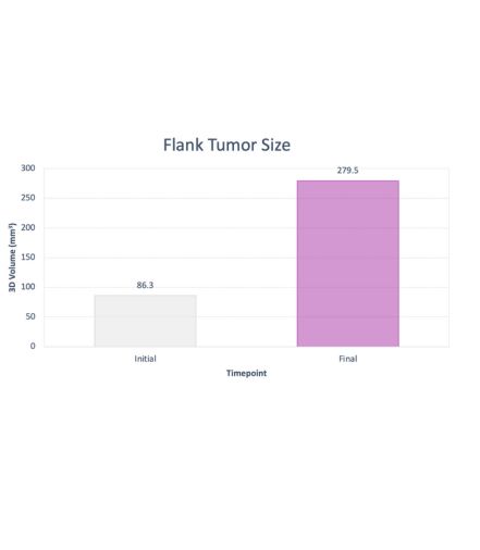
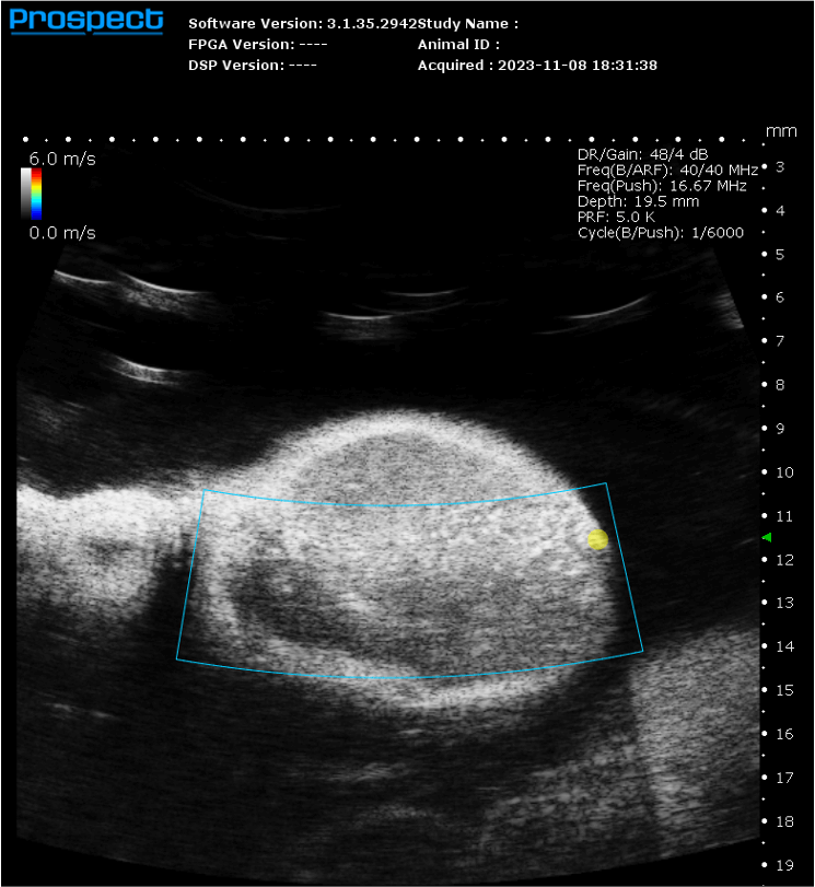
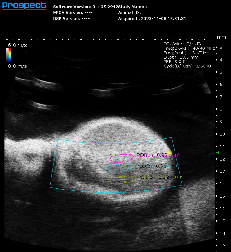
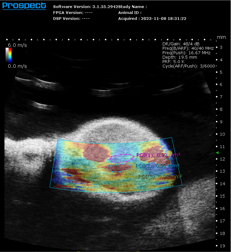
- Oncology
- Neurology
- Developmental Biology
- Abdominal Anatomy
- Pulmonary Biology
- Musculoskeletal Anatomy
- Drug Discovery
- Safety Pharmacology
- Microbiology & Immunology
- Regenerative Medicine
- Bioprinter
- Engineering & System Components
- Research/Therapeutic US
- Data Acquisition and Preamplification Electronics
- Engineering PA/US
- Other Animal Models
- Services
- Resources
- About Us
- Shop
- Contact Us


