Pursuing more accurate, detailed, and less invasive imaging systems is unending in the dynamic realm of preclinical research. Enter BLI/FLI techniques, the cutting-edge of optical imaging that is reshaping the biomedical research landscape. These tools have emerged as cornerstones of preclinical study, offering unparalleled real-time insights into in vivo biological processes. But what sets BLI/FLI techniques apart from traditional imaging modalities?
Optical Imaging
At their core, BLI/FLI techniques represent optical imaging’s finest, harnessing the power of light to unveil intricate biological processes. Distinct from the limitations of conventional imaging, these tools present a synthesis of clarity and depth, extending the boundaries of preclinical investigations.
Bioluminescence Imaging (BLI)
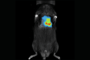
BLI leverages the natural light emitted by living organisms. This process is orchestrated by the luciferase enzymes, which facilitate the oxidation of luciferin, culminating in light production. The emitted glow translates to a visual representation of myriad biological phenomena. It is a game-changer in preclinical imaging because it allows researchers to track and quantify biological processes in vivo with high sensitivity and specificity.
BLI has been used to study a wide range of biological processes:
- Fungal infections[1]
- Neutrophil tracking[2]
- Lung metastasis detection[3]
- Cancer progression[4]
It also has broad applicability in preclinical drug studies[5]. It is particularly useful in preclinical drug development as it enables researchers to monitor drug efficacy in real-time. This is a boon for drug dosing optimization and administration scheduling. BLI is also extremely valuable in developing fluorescence-guided surgery and patient-derived orthotopic mouse models[5].
Fluorescence Imaging (FLI)
FLI differs from BLI in that it uses fluorescent molecules to unveil biological intricacies. These molecules emit a discernible light when excited by a specific wavelength. This luminescence offers a real-time depiction of biological processes unfettered by invasive techniques. It is another non-invasive imaging technique that measures the lifetime of fluorescent molecules to visualize biological processes in real time. This makes FLI a powerful tool for studying metabolic changes in cells and tissues. It has has been used to study a wide range of biological processes, including:
- Cancer progression[6]
- Protein-protein interactions[7]
- Mitochondrial function[8].
A fundamental advantage of FLI is that it can provide quantitative information about the concentration and distribution of fluorescent molecules in cells and tissues. It can also distinguish between different fluorescent molecules based on their lifetime, allowing for multiplexing and the simultaneous detection of multiple targets.
Advantages: Why BLI/FLI Triumphs
Beyond their foundational differences, BLI/FLI techniques present several operational advantages. They eclipse traditional modalities like X-ray imaging, proffering three-dimensional, high-resolution insights sans radiation-associated risks. Furthermore, these techniques unveil biological processes in vivo, granting researchers a reflective view of disease progression, therapeutic responses, and tissue regeneration.
Applications: The Broad Spectrum
The versatility of BLI/FLI is evident in its vast applicability, spanning from plant imaging to intravital microscopy. Preclinical imaging, especially, stands as a testament to its prowess. By facilitating a closer look at gene expression, protein-protein interactions, and cellular signaling, these techniques furnish researchers with a more holistic understanding of living organisms.
In essence, BLI/FLI techniques serve as optical imaging’s zenith, employing light as a medium to demystify complex biological processes. With advantages that dwarf traditional methodologies and a versatility that caters to diverse research needs, they indubitably lead the way in modern biomedical research.
Embracing the Future Preclinical Optical Imaging with Scintica
At Scintica, we recognize the transformative potential of BLI/FLI techniques. We’re not just observers but pioneers dedicated to pushing the envelope in preclinical research. For those poised to redefine the boundaries of scientific inquiry, we invite you to explore our cutting-edge optical imaging systems. Dive deep, innovate, and let’s shape the future of biomedical research together.
References and further reading:
- Gago, S., Denning, D. W., Bowyer, P., & Bromley, M. J. (2020). Powerful and Real-Time Quantification of Antifungal Efficacy against Triazole-Resistant and -Susceptible Aspergillus fumigatus Infections in Galleria mellonella by Longitudinal Bioluminescence Imaging. Frontiers in microbiology, 11, 573. doi: 10.3389/fmicb.2020.00573
- Chalishazar, M. D., & Kimmelman, A. C. (2022). Abstract 2455: Development of a bioluminescence reporter mouse model for tracking and quantifying Ly6G+ neutrophils in vivo. Cancer Research, 82(13 Supplement), 2455. doi: 10.1158/1538-7445.AM2022-2455
- Van de Wiele, C., Vanhove, C., & Lahoutte, T. (2017). Longitudinal microcomputed tomography-derived biomarkers for lung metastasis detection in a syngeneic mouse model: added value to bioluminescence imaging. Molecular imaging, 16, 1536012116679653. doi: 10.1177/1536012116679653
- Piwnica-Worms D. From the Guest Editor: Illuminating Cancer In Vivo With Molecular Imaging. Cancer J. 2015 May-Jun;21(3):150-1. doi: 10.1097/PPO.0000000000000121. PMID: 26049693.
- Irtenkauf SM, Sobiechowski S, Hasselbach LA, Nelson KK, Transou AD, Carlton ET, Mikkelsen T, deCarvalho AC. Optimization of Glioblastoma Mouse Orthotopic Xenograft Models for Translational Research. Comp Med. 2017 Aug 1;67(4):300-314. PMID: 28830577; PMCID: PMC5557202.
- Kostoff, R. N., & Barth, R. (2015). References for scientific papers: why not standardise to one global style?. F1000Research, 4, 28. doi: 10.12688/f1000research.4-28.v1
- Kwon, S., & Kim, S. (2016). Bioluminescence imaging in vivo for stem cell research and therapy. International journal of molecular sciences, 17(6), 826. doi: 10.3390/ijms17060826
- Zhang, X., & Li, Y. (2021). Bioluminescence Imaging of Tumor-Associated Macrophages in Cancer Immunotherapy. Frontiers in immunology, 12, 661758. doi: 10.3389/fimmu.2021.661758