(February 29, 2024) Webinar: In Vivo Photoacoustic Tomography in Assessing Acute Systemic Vasoactivity: A Noninvasive Systemic Myography Tool
Overview:
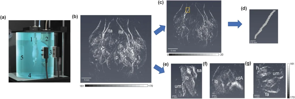
In this webinar, Dr. Kristie Huda demonstrated how photoacoustic tomography can be used as a noninvasive in vivo myography technique to evaluate systemic vascular reactivity of pregnant mice in response to targeted therapeutics. An alternate method to traditional vessel myography that requires the isolation of individual arteries to assess the vascular response, photoacoustic tomography opens a new gateway to extract invaluable preclinical information of whole-body vascular response to therapeutics that is not possible with conventional myography techniques.
Altered vascular reactivity is a critical indicator of cardiometabolic diseases such as hypertension, diabetes, atherosclerosis, coronary artery disease, and renal diseases. Preclinical measurement of vascular reactivity to therapeutics is an effective method to determine the efficacy of therapeutics in the treatment of cardiometabolic diseases. Selective therapeutics are ideal to maximize the benefit of the treatment as well as avoiding side effects like systemic hypotension. Conventional myography techniques require the excision of a large, individual artery (wire myography), surgically exposed tissue (Intravital microscopy), or superficial single artery (noninvasive 2D ultrasound imaging) to assess and quantify vascular response. However, these techniques may not represent the in vivo physiological condition accurately due to the excision of an artery and in vivo vascular changes in 3D space.
Photoacoustic tomography captures the whole-body anatomical and functional information of a small animal in a 3D volumetric image. Using the TriTom photoacoustic tomography system, they sought to assess vascular reactivity both systemically and in the placenta during pregnancy. Their study assessed sildenafil and G-1 (a selective agonist of the G protein-coupled estrogen receptor) vasodilators with different pathways to induce vascular reactivity in systemic vasculature and placenta.
Key Takeaways:
- Photoacoustic tomography enables visualization of systemic arteries of maternal vasculature, placentas, and fetal vasculature.
- Photoacoustic tomography captures acute vasodilation of major arteries and detects selective vasoactivity in maternal-fetal vasculature.
- TriTom and the developed method in this study can be applied to test novel therapeutics on an animal model to study their systemic and off-target side effects.
Disclaimer:
The research presented in this webinar was conducted at Tulane University prior to employment at GE HealthCare.
Huda, K., Lawrence, D.J., Thompson, W. et al. In vivo noninvasive systemic myography of acute systemic vasoactivity in female pregnant mice. Nat Commun 14, 6286 (2023). https://doi.org/10.1038/s41467-023-42041-8

About the Speaker (s)

Dr. Kristie Huda,
Research Scientist at GE HealthCare Technology & Innovation Center
Dr. Kristie Huda is a Research Scientist in the Ultrasound and Bioinstrumentation group at GE HealthCare Technology and Innovation Center, Niskayuna, NY. She completed her Ph.D. in Biomedical Engineering from Tulane University, New Orleans, LA under the supervision of Dr. Carolyn Bayer. Her dissertation focused on developing methods for photoacoustic tomography to assess systemic functional and molecular changes in vivo and demonstrate the clinical applicability of photoacoustic transabdominal imaging for humans through simulation. Kristie is passionate about women’s health research. She is motivated to find novel imaging solutions to challenging medical imaging problems.