System Used:
IVIM System

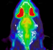
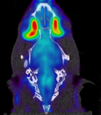
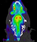
Advancements in imaging techniques have revolutionized biomedical research by enabling scientists to visualize and study biological processes at various scales. While whole-body imaging modalities, such as magnetic resonance imaging (MRI), ultrasound, bioluminescence and fluorescence imaging, and positron emission tomography (PET), provide valuable macroscopic information, however, they are limited in their ability to capture cellular and subcellular dynamics.
PET/CT
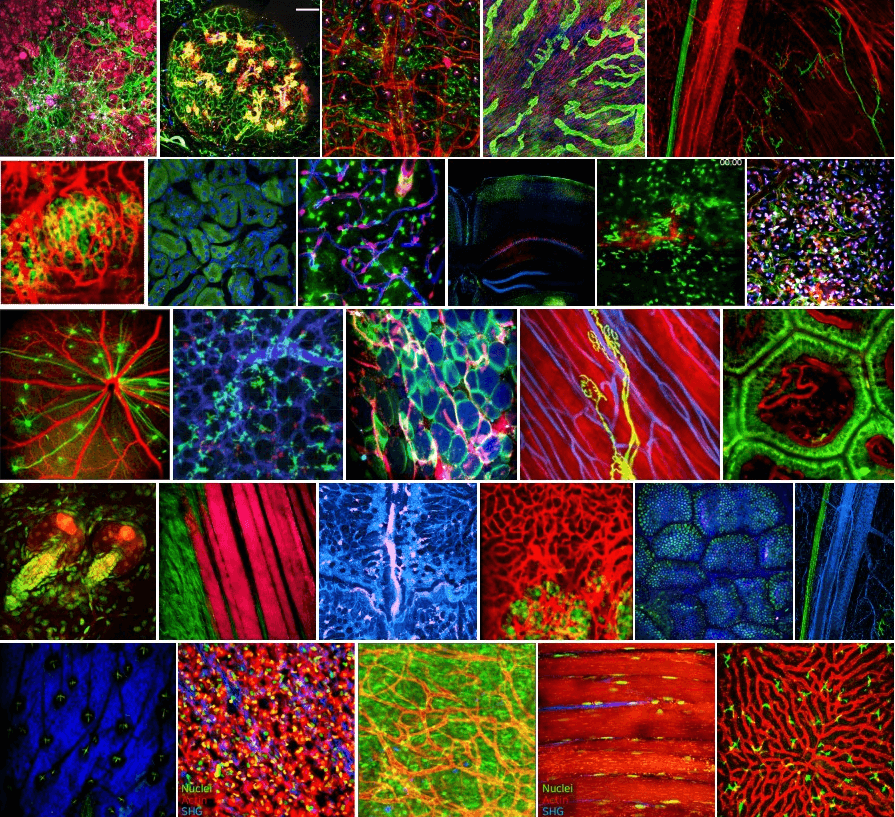
Intravital microscopy, on the other hand, offers high-resolution imaging at the cellular and subcellular level in living organisms, providing unique insights into dynamic processes within specific tissue microenvironments. This article aims to explore the importance of integrating intravital microscopy with whole-body imaging modalities, highlighting the complementary nature of these techniques and their contributions to advancing biomedical research.
Whole-body imaging modalities are valuable for capturing macroscopic changes in anatomy, physiology, and metabolism. However, to fully comprehend the underlying mechanisms of disease and physiological processes, it is essential to investigate cellular and subcellular dynamics. Intravital microscopy excels in providing real-time visualization of cellular events, such as cell migration, immune responses, and cellular interactions within tissues. By combining intravital microscopy with whole-body imaging techniques, researchers can bridge the gap between macroscopic observations and microscopic insights, facilitating a comprehensive understanding of complex biological processes.
One of the significant advantages of intravital microscopy is its ability to capture dynamic processes in real-time. Unlike whole-body imaging modalities that often provide static or averaged data, intravital microscopy allows researchers to observe cellular behavior and interactions as they occur. This real-time imaging is particularly crucial when studying dynamic processes, such as immune cell recruitment, tumor progression, and vascular dynamics. Moreover, intravital microscopy enables longitudinal studies, allowing researchers to monitor the same region of interest over time, providing valuable insights into disease progression, treatment response, and tissue regeneration.
Intravital microscopy provides precise localization of cellular and molecular events within specific tissue microenvironments. This spatial context is essential for understanding the interactions between cells, extracellular matrix components, vasculature, and immune cells. By visualizing these interactions at the microscopic scale, researchers can gain insights into disease progression, immune responses, and drug efficacy. Whole-body imaging techniques may provide information about the distribution of tracers or contrast agents, but they lack the ability to assess cellular and molecular interactions within specific tissue niches. Integrating intravital microscopy with whole-body imaging provides a comprehensive understanding of how cellular behavior and tissue microenvironments contribute to overall physiological changes.
Intravital Microscopy
While whole-body imaging modalities are excellent for assessing global physiological and anatomical changes, intravital microscopy offers detailed functional and structural information at the local level. Intravital microscopy allows researchers to measure parameters such as blood flow, oxygenation, calcium signaling, and membrane potential in real-time, providing insights into tissue and organ function. By combining functional measurements obtained through intravital microscopy with anatomical whole-body imaging data, researchers can correlate global changes with localized cellular and subcellular events, leading to a more comprehensive understanding of disease processes and treatment responses.
Integrating intravital microscopy with whole-body imaging modalities offers a powerful tool for validating and further understanding macroscopic observations. Whole-body imaging techniques generate macroscopic data that can be challenging to interpret without detailed knowledge of the underlying cellular and molecular events. Intravital microscopy allows researchers to directly visualize and validate the presence or absence of specific cellular markers, disease phenotypes, or therapeutic responses observed in whole-body imaging studies. This validation enhances the reliability and interpretation of whole-body imaging data, reinforcing the link between microscopic and macroscopic observations.
The integration of intravital microscopy with whole-body imaging modalities is a rapidly evolving field, and ongoing advancements are opening up new possibilities for comprehensive biomedical research. Technological innovations, such as multiphoton microscopy, adaptive optics, and genetically encoded fluorescent probes, are enhancing the capabilities of intravital microscopy, enabling deeper tissue penetration, improved resolution, and selective imaging of specific cell populations. Furthermore, advancements in image analysis and data integration techniques are facilitating the integration of data obtained from different imaging modalities, enabling researchers to extract more comprehensive and meaningful insights from multimodal imaging experiments.
The integration of intravital microscopy with whole-body imaging modalities is instrumental in advancing our understanding of complex biological processes, disease mechanisms, and treatment responses. While whole-body imaging provides valuable macroscopic information, intravital microscopy offers the unique ability to visualize cellular and subcellular dynamics in living organisms. By combining these techniques, researchers can bridge the gap between macroscopic observations and microscopic insights, facilitating a comprehensive understanding of disease progression, tissue microenvironments, and therapeutic interventions. As technology continues to advance, the integration of these imaging modalities will play an increasingly critical role in unraveling the complexities of biomedical research and improving healthcare outcomes.
Optical Imaging