Optical imaging has become an indispensable tool in drug development. This technology, which includes bioluminescence imaging, fluorescence imaging (FLI), and near-infrared imaging (NIR), has significantly enriched our capacity to perform thorough biodistribution and molecular imaging studies, thereby facilitating a smoother transition from concept to clinic.
Biodistribution Studies: A Pillar in Drug Development
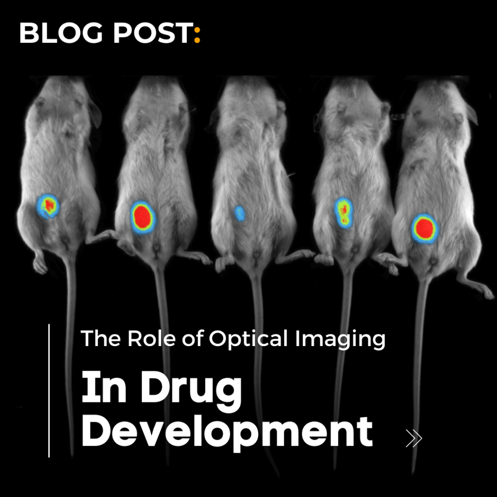
Biodistribution studies, leveraging near-infrared optical imaging, have become crucial in observing and analyzing the dynamic distribution of drugs within living organisms. The accuracy and dynamism of optical imaging enable the tracking of drugs, providing researchers with insights into how substances navigate through various biological compartments, and their interactions with targeted and non-targeted tissues. Understanding the pharmacokinetics and pharmacodynamics of the drug ensures it reaches the intended target and exerts the desired therapeutic effect while minimizing off-target interactions.
Molecular Imaging Studies: A Microscopic View
In molecular imaging studies, optical imaging provides a detailed view, allowing scientists to visualize molecular events in living organisms, such as the expression of reporter genes. This capability is vital in evaluating the molecular mechanisms through which drugs exert their effects and identifying potential biomarkers for disease progression and therapeutic response. By providing a window into the cellular and molecular level, optical imaging enables researchers to scrutinize the intricate interactions between drugs and biological pathways, thereby facilitating the development of more targeted and effective therapeutic agents.
Preclinical Imaging: Bridging the Gap to Clinical Trials
Preclinical imaging, especially with systems like the Newton 7.0, which integrates optical bioluminescence and 3D tomographic imaging, has been instrumental in preclinical research endeavors such as drug development, disease modeling, and molecular imaging studies in small animals. The Newton 7.0 provides a robust platform for conducting comprehensive preclinical studies, offering insights that are pivotal in shaping subsequent clinical trials and ensuring that potential therapeutic candidates have a solid foundation of efficacy and safety data before progressing to human studies.
Non-Invasive Imaging: Ensuring Safety and Detail
The non-invasive nature of optical imaging ensures that detailed images of internal structures can be obtained without subjecting organisms to the potential risks associated with radiation.
This is not only crucial in safeguarding the well-being of the subjects but also in ensuring that the deleterious effects of invasive procedures or radiation exposure do not confound the data obtained. The ability to conduct repeated measurements in a safe manner also allows for longitudinal studies, where the effects of drugs can be monitored over extended periods, providing a more comprehensive understanding of their long-term efficacy and safety.
Scintica’s Vision for the Future
At Scintica, we have witnessed and facilitated the remarkable advancements in drug development brought about by optical imaging. From conducting intricate biodistribution studies to enabling detailed molecular imaging studies, optical imaging has proven to be an invaluable tool in bridging the gap from concept to clinic.
The Newton 7.0, with its capabilities in optical bioluminescence and 3D tomographic imaging, represents our commitment to furthering this field, providing researchers with the tools they need to delve deeper into the mysteries of drug interactions and biological systems. We invite you to explore the possibilities unlocked by the Newton 7.0 and join us in paving the way towards a future where the journey from concept to clinic is streamlined, efficient, and above all, illuminated by the unparalleled insights provided by optical imaging. Discover more about the Newton 7.0.