 In this journal article published in early February 2020 titled “Sight and switch off: Nerve density visualization for interventions targeting nerves in prostate cancer” in Science Advances, Huijuan You et al. discuss the development of their novel superparamagnetic iron oxide nerve peptide nanoparticles (PSN NPs) to image and quantify nerve density in a mouse model of prostate cancer using the M3 compact MRI system from Aspect Imaging. This group took the next step to show the therapeutic capabilities of this compound with targeted drug delivery to the nerves to inhibit tumor progression.
Nerve density has been shown to be associated with the prognosis of prostate cancer patients. When nerve density is high, tumors progress and the prognosis are poor, however, when nerve density is low the outcomes are better. Having the ability to visualize and quantify nerve density in the clinic would allow physicians to provide personalized medicine (i.e. to treat appropriately based on the specific tumor properties) that would help prevent overtreatment in low nerve density tumors, but would allow appropriately high levels of treatment in those with higher nerve density. Adding targeted drug delivery to these nanoparticles empowers them as a theranostic compound, that is both diagnostic and therapeutic.
In this journal article published in early February 2020 titled “Sight and switch off: Nerve density visualization for interventions targeting nerves in prostate cancer” in Science Advances, Huijuan You et al. discuss the development of their novel superparamagnetic iron oxide nerve peptide nanoparticles (PSN NPs) to image and quantify nerve density in a mouse model of prostate cancer using the M3 compact MRI system from Aspect Imaging. This group took the next step to show the therapeutic capabilities of this compound with targeted drug delivery to the nerves to inhibit tumor progression.
Nerve density has been shown to be associated with the prognosis of prostate cancer patients. When nerve density is high, tumors progress and the prognosis are poor, however, when nerve density is low the outcomes are better. Having the ability to visualize and quantify nerve density in the clinic would allow physicians to provide personalized medicine (i.e. to treat appropriately based on the specific tumor properties) that would help prevent overtreatment in low nerve density tumors, but would allow appropriately high levels of treatment in those with higher nerve density. Adding targeted drug delivery to these nanoparticles empowers them as a theranostic compound, that is both diagnostic and therapeutic.
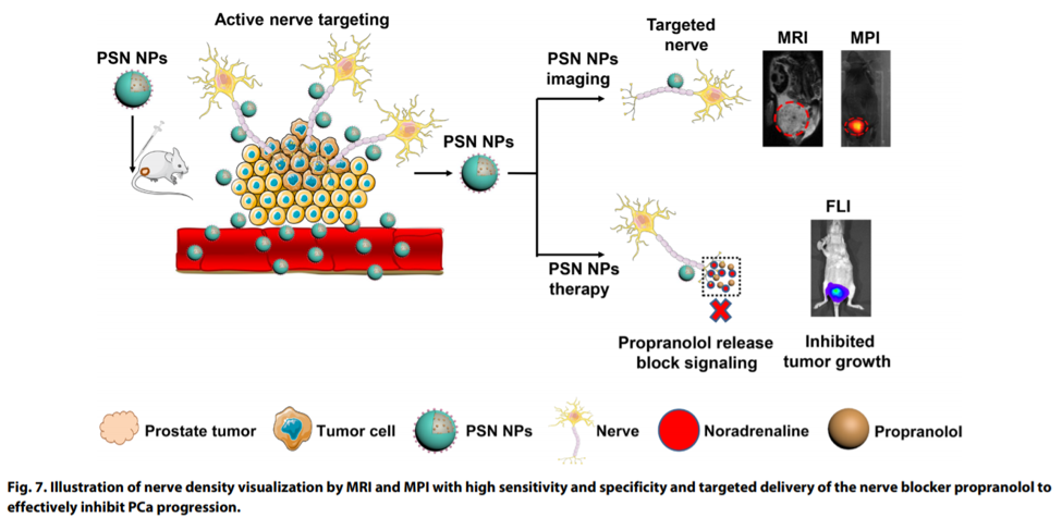
 In this study, PC-3luc cells were inoculated into the ventral prostate of BALb/c nude male mice. This group also used a variety of other xenogeneic prostate tumor models having known differences in nerve density. This allowed the group to study tumor progression with bioluminescence along with MRI imaging. Nanoparticles were synthesized to either bind to nerves (PSN NPs) or not (PS NPs). As they are superparamagnetic iron oxide nanoparticles, they are visible on a T2 weighted MR image as a drop out of signal. Likewise, these same nanoparticles can also be detected using a newer technology called magnetic particle imaging (MPI), which was utilized by this group as a technology in their studies.
You et al. report that they were able to develop and use these nanoparticles to visualize nerve density in their tumor model. Additionally, with the targeted drug delivery they were able to inhibit the progression of prostate tumors in their mouse model. Quantification of nerve density, as measured by MRI, correlated well with the MPI results which were both validated against histological results as the gold standard. Bioluminescence was also used to monitor tumor progression, especially in the treated study groups.
Please reach out to us at Scintica Instrumentation to learn more about the Aspect Imaging M-Series MRI systems, and our bioluminescence imaging systems.
In this study, PC-3luc cells were inoculated into the ventral prostate of BALb/c nude male mice. This group also used a variety of other xenogeneic prostate tumor models having known differences in nerve density. This allowed the group to study tumor progression with bioluminescence along with MRI imaging. Nanoparticles were synthesized to either bind to nerves (PSN NPs) or not (PS NPs). As they are superparamagnetic iron oxide nanoparticles, they are visible on a T2 weighted MR image as a drop out of signal. Likewise, these same nanoparticles can also be detected using a newer technology called magnetic particle imaging (MPI), which was utilized by this group as a technology in their studies.
You et al. report that they were able to develop and use these nanoparticles to visualize nerve density in their tumor model. Additionally, with the targeted drug delivery they were able to inhibit the progression of prostate tumors in their mouse model. Quantification of nerve density, as measured by MRI, correlated well with the MPI results which were both validated against histological results as the gold standard. Bioluminescence was also used to monitor tumor progression, especially in the treated study groups.
Please reach out to us at Scintica Instrumentation to learn more about the Aspect Imaging M-Series MRI systems, and our bioluminescence imaging systems.