Our cutting-edge in vivo imaging technology now enables long-term imaging of astrocytes and microglia in the hippocampus and hypothalamus – deep within the brain – using precise surgical animal modeling methods.
We’ve enhanced existing window chamber and implantation techniques to gain deeper insights into drug reaction mechanisms in degenerative brain diseases, aligning perfectly with the current trends in research. In contrast to conventional methods primarily focused on the cortex and meninges, our precision surgery approach minimizes bleeding and side effects while providing a more authentic view of deep brain tissue. This facilitates in vivo environment imaging at cellular to subcellular resolutions.
In this newsletter, you will have the chance to explore the revolutionary Cranial Imaging Cannula (CIC) surgical method, meticulously designed by IVIM Technology. It enables the imaging of neural activity within the hippocampus and hypothalamus—both located deep within the brain tissue. Additionally, we’ll introduce IVIM’s Needle Type Endomicroscopy and imaging technique, specifically tailored to capture neural activity in the depths of the brain.
Lastly, you will be introduced to our Optical Clearing imaging technology, a solution that eradicates unnecessary tissue interference, ensuring clear and precise imaging by minimizing laser scattering.
I. Cranial Imaging Cannula (CIC): Precision Post-Surgical Imaging Technology for Visualizing Hippocampal and Hypothalamic Nerve Activity
The Cranial Imaging Cannula (CIC) is a cannula insertion surgery and imaging technique designed for real-time visualization of hippocampal neural activity and microglial cells within living tissues. This technique has been optimized to visualize neural activity within the hippocampus, located deep in the brain tissue. It serves as an invaluable imaging tool, for longitudinal imaging of the same region of interest within the same subject over the course of several weeks/months.
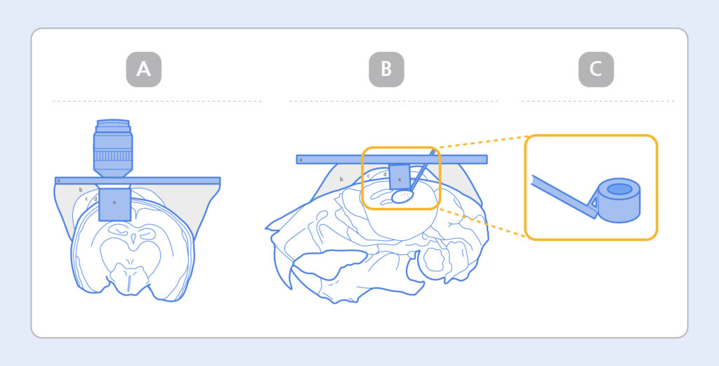
Picture 1. Diagram of A) Microscope mounting photo of the brain tissue after the Cranial Imaging Cannula (CIC) implantation, B) CIC implantation in the upper part of hippocampus CA1 region, C) Cranial Imaging Cannula.
In our pursuit of establishing a comprehensive protocol for imaging hippocampal and hypothalamic neural activity using the CIC method, IVIM`s dedicated research team has constructed a biomicroscopy module tailored to the unique requirements of CIC experiments. (Pic. 1) This module integrates all essential components, from CIC experiment tools to brain tissue mounting platforms.
For imaging deep regions of brain tissue utilizing the CIC technique, we employ the IVM-CM3, specifically designed for two-photon imaging. This powerful tool enables imaging of neural tissue activity within the hippocampus and hypothalamus at a cellular level.
II. Intravital Imaging Leveraging Needle Type Endomicroscopy for Precise Capture of Neural Activity at Various Depths within the Brain
Needle-type endomicroscopy is a cutting-edge imaging technology designed for real-time visualization of neural activity at various depths within living brain tissue. This groundbreaking technology facilitates imaging at distinct brain tissue depths including critical regions such as the Cortex, Hippocampus, and Thalamus.
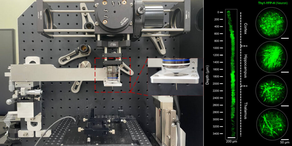
Picture 2. Needle Type Endomicroscopy (Left) and Imaging Result (Right)
Through the optimization of bioimaging techniques utilizing Needle-Type Endomicroscopy, our research team has achieved depth-specific brain tissue imaging. After creating a 3mm craniotomy in the area of Bregma, needle-type endomicroscopy is performed without a need for optical clearing. Images of brain tissue sections in the Cortex, Hippocampus, and Thalamus of a Thy-1-YFP-16 transgenic were acquired.
III. Intravital Imaging Combined with Optical Clearing Solution: Studying the Neural Activity Depp within the Brain Tissue
Another method for imaging deep regions within the brain involves the use of Optical Clearing techniques. Optical clearing makes biological tissues transparent. Living tissues consist of various components, including fat, which can scatter laser wavelengths and hinder clear imaging using a biological microscope
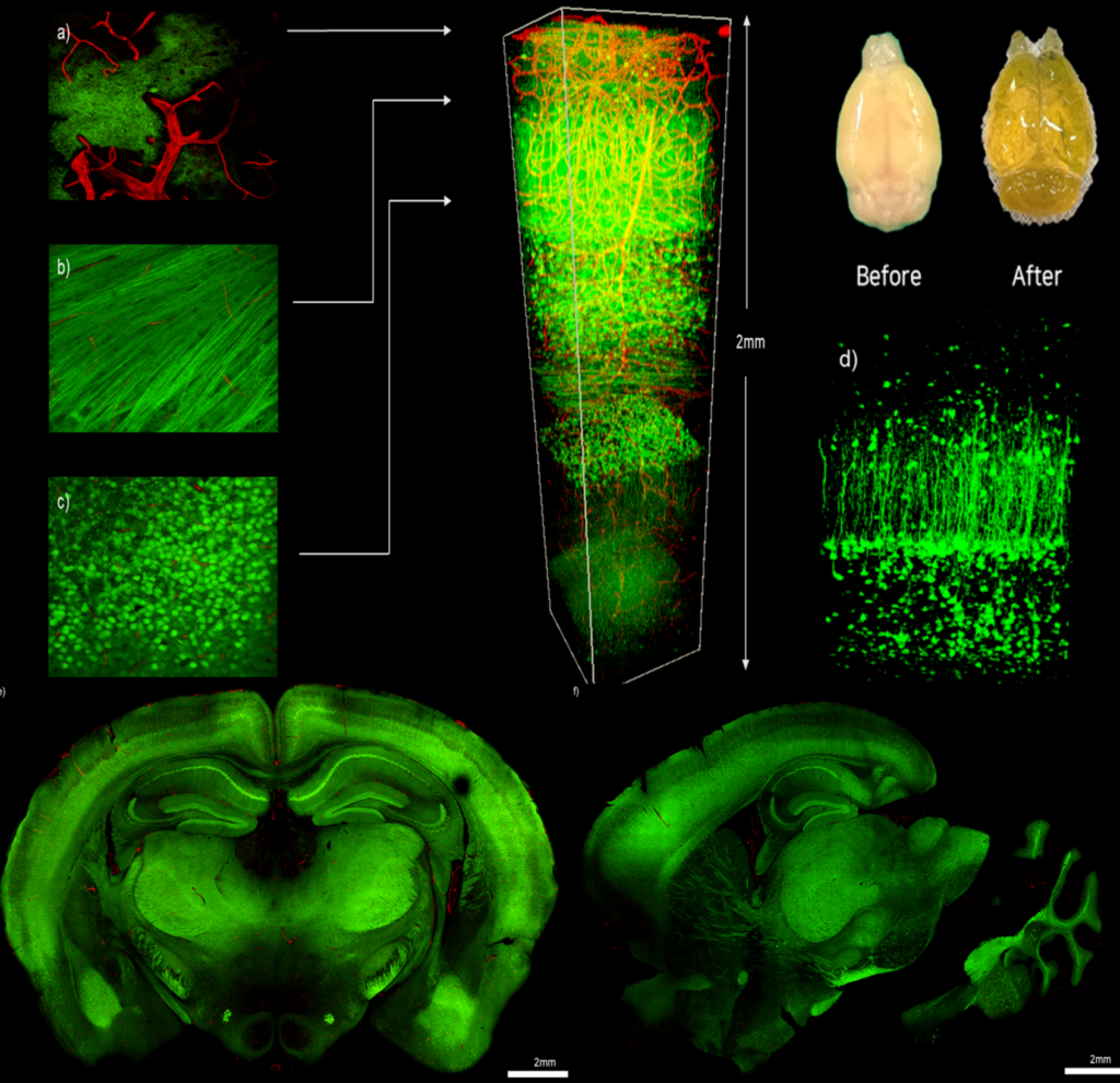
Picture 3. Fluorescence Imaging of Thy1-YFP (H Line) Mouse Brain at the a) Brain Surface Layer (Pial Surface), b) Alveus Layer or White Matter, and c) Hippocampus Pyramidal Cells. d) Fluorescence Imaging of YFP-Expressing Pyramidal Neurons in 3D Zoom Image. e, f) Confocal Coronial View of a Thy1-YFP-16 Brain Slice.
The IVIM research team conducted brain extractions from live animals to delve into the inner workings of brain tissue, utilizing Thy-1-YFP-16 mice—a transgenic mouse model featuring neural tissue markers. Leveraging the IVM-CM3, a state-of-the-art system equipped with two-photon mode imaging and high-speed real-time laser scanning capabilities, they explored the potential of imaging the brain from various angles and depths, spanning from its surface to the most profound regions.
Furthermore, by harnessing the Z-axis cross-sectional imaging function, they were able to capture images as deep as 2 millimeters within the brain tissue using two-photon mode microscopy.
This breakthrough opens new avenues for in-depth exploration of the brain’s internal mechanisms.