Invicro and Scintica Instrumentation have teamed up to help solve a fundamental issue of microscopic imaging; quantification can be difficult without customizable image analysis software.
The ViewnVivo confocal endomicroscope is used to obtain clear, high quality in vivo images of tissues. This enables users to qualitatively interpret and compare these images.
Of course, any manual human based analysis is subject to interpreter bias and differing explanations from other researchers. For this reason, many journals are hesitant to publish microscopic images alone and require additional data. This constraint has constantly made in vivo microscopy a secondary technique supporting other lab-based methods.
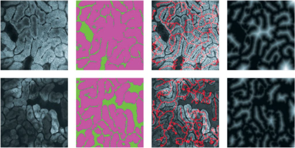
Invicro is the first group to work with the ViewnVivo to establish a technique to obtain endoscopic confocal images and generate quantitative data. Through their custom image analysis algorithms, Invicro explored, analyzed, and compared various normal and abnormal rodent tissues.
The White Paper below details Invicro’s analysis of liver, kidney, colon and blood vessels (colon and brain) and their approach to creating quantifiable data on which informative statistics can be acquired. Obtaining quantifiable data from images should help confocal endomicroscopy become a standard microscopic technique for more in vivo based publications.
- Products
- Imaging
- Microscopy
- Bioprinting
- Lab Equipment
- Surgery
- Physiology
- Cell & Isolated Tissue
- Hypoxia & Atmospheric Control
- Data Acquisition Solutions
- Software
- Phantoms
- Consumables
- Applications
- Cardiovascular Biology
- Oncology
- Neurology
- Developmental Biology
- Abdominal Anatomy
- Pulmonary Biology
- Musculoskeletal Anatomy
- Drug Discovery
- Safety Pharmacology
- Microbiology & Immunology
- Regenerative Medicine
- Bioprinter
- Engineering & System Components
- Research/Therapeutic US
- Data Acquisition and Preamplification Electronics
- Engineering PA/US
- Other Animal Models
- Services
- Resources
- About Us
- Shop
- Contact Us