
In Situ X-Ray-Induced Acoustic Computed Tomography With a Contrast Agent: A Proof of Concept
Authors: Seongwook Choi1; Sinyoung Park1; Ayoung Pyo2; Dong-Yeon Kim3 ; Jung-Joon Min4 ; Changho Lee,4,5,6; Chulhong Kim1,7
System Used:
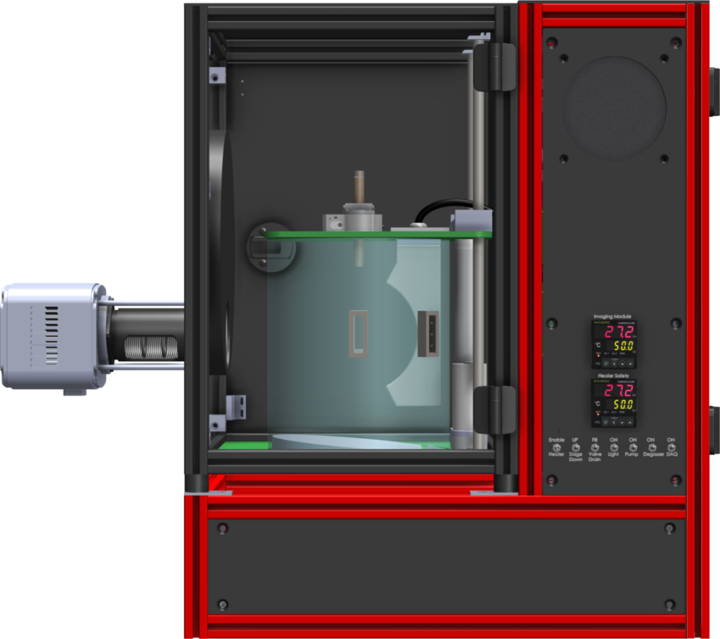
TRITOM
AFFILIATIONS
1 Department of Electrical Engineering, Convergence IT Engineering, and Mechanical Engineering, Medical Device Innovation Center, Pohang
University of Science and Technology, Pohang, Republic of Korea
2 Korea Atomic Energy Research Institute, Jeongeup, Republic of Korea
3 College of Pharmacy and Research Institute of Pharmaceutical Science, Gyeongsang National University, Jinju, Republic of Korea
4 Department of Nuclear Medicine, Chonnam National University Medical School & Hwasun Hospital, Hwasun, Chonnam, Republic of Korea
5 Department of Artificial Intelligence Convergence, Chonnam National University, Gwangju, Republic of Korea
6 e-mail: ch31037@jnu.ac.kr
7 e-mail: chulhong@postech.edu
ABSTRACT
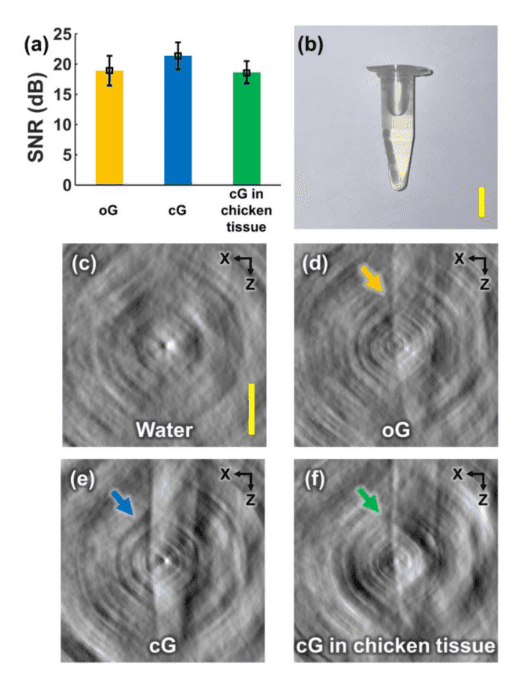
X-ray-induced acoustic computed tomography (XACT) has shown great potential as a hybrid imaging modality for real-time non-invasive x-ray dosimetry and low-dose three- dimensional (3D) imaging. While promising, one drawback of the XACT system is the underlying low signal-to-noise ratio (SNR), limiting its in vivo clinical use. In this Letter, we propose the first use of a conventional x-ray computed tomography contrast agent, Gastrografin, for improving the SNR of in situ XACT imaging. We obtained 3D volumetric XACT images of a mouse’s stomach with orally injected Gastrografin establishing the proposal’s feasibility. Thus, we believe, in the future, our proposed technique will allow in vivo imaging and expand or complement conventional x-ray modalities, such as radiotherapy and accelerators.
REFERENCE
1. L. Xiang, B. Han, C. Carpenter, G. Pratx, Y. Kuang, and L. Xing, Med. Phys. 40, 010701 (2012).
2. S. Hickling, M. Hobson, and I. El Naqa, Int. J. Radiat. Oncol., Biol., Phys. 90, S843 (2014).
3. S. Hickling, P. Léger, and I. El Naqa, IEEE Trans. Ultrason., Ferro-elect., Freq. Contr. 63, 683 (2016).
4. L. Xiang, S. Tang, M. Ahmad, and L. Xing, Sci. Rep. 6, 26118 (2016).
5. J. Kim, E.-Y. Park, Y. Jung, B. C. Kim, J. H. Kim, C.-Y. Yi, I. J. Kim, and C. Kim, IEEE Trans. Radiat. Plasma Med. Sci. 1, 534 (2017).
6. S. Tang, D. Nguyen, A. Zarafshani, C. Ramseyer, B. Zheng, H. Liu, and L. Xiang, Appl. Phys. Lett. 110, 103504 (2017).
7. S. Tang, C. Ramseyer, P. Samant, and L. Xiang, Appl. Phys. Lett. 112, 063504 (2018).
8. S. Tang, K. Yang, Y. Chen, and L. Xiang, Med. Phys. 45, 1662 (2018).
9. H. Lei, W. Zhang, I. Oraiqat, Z. Liu, J. Ni, X. Wang, and I. El Naqa, Med. Phys. 45, 4191 (2018).
10. F. Forghani, A. Mahl, T. J. Patton, B. L. Jones, M. A. Borden, D. C. Westerly, C. Altunbas, M. Miften, and D. H. Thomas, Med. Phys. 47, 1280 (2020).
11. W. Zhang, I. Oraiqat, H. Lei, P. L. Carson, I. EI Naqa, and X. Wang, BME Frontiers 2020, 9853609 (2020).
12. Y. Li, P. Samant, S. Wang, A. Behrooz, D. Li, and L. Xiang, IEEE Trans. Ultrason., Ferroelect., Freq. Contr. 67, 1613 (2020).
13. E. Robertson, P. Samant, S. Wang, T. Tran, X. Ji, and L. Xiang, IEEE Trans. Ultrason., Ferroelect., Freq. Contr. 68, 1073 (2021).
14. P. Samant, L. Trevisi, X. Ji, and L. Xiang, J. Photoacoust. 19, 100177 (2020).
15. S. Choi, E.-Y. Park, S. Park, J. H. Kim, and C. Kim, Sci. Rep. 11, 1 (2021).
16. D. Lee, E.-Y. Park, S. Choi, H. Kim, J.-j. Min, C. Lee, and C. Kim, Biomed. Opt. Express 11, 752 (2020).
17. S. Choi, D. Lee, E.-Y. Park, J.-J. Min, C. Lee, and C. Kim, in Photons Plus Ultrasound: Imaging and Sensing 2020, (International Society for Optics and Photonics, 2020), 112404R.
18. S. Cho, J. Baik, R. Managuli, and C. Kim, J. Photoacoust. 18, 100168 (2020).