System Used In This Work:
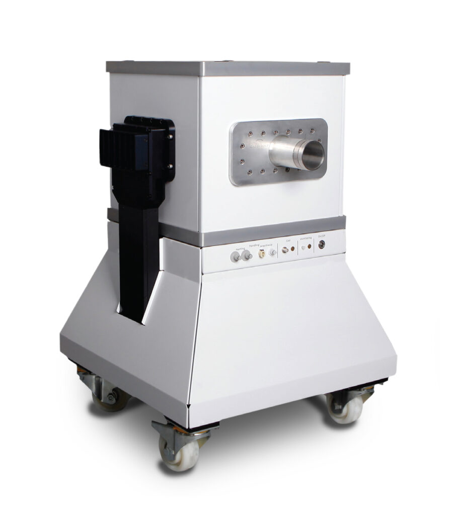
M-Series MRI
Sharma, G.; Braga, M.C.; Da Pieve, C.; Szopa, W.; Starzetz, T.; Plate, K.H.; Kaspera, W.; Kramer-Marek, G. Immuno-PET Imaging of Tumour PD-L1 Expression in Glioblastoma. Cancers 2023, 15, 3131. https://doi.org/10.3390/cancers15123131
The prognosis of patients with Glioblastoma (GBM), a highly aggressive malignant brain tumor, remains extremely poor due to a lack of effective therapeutic options. Since it was voted the ‘breakthrough of the year’ by Science in 2013, immunotherapy has revolutionized the treatment of solid tumors. This includes small-molecule immune checkpoint inhibitors targeting either the programmed cell death protein 1 (PD-1, e.g., nivolumab, pembrolizumab) or its ligand (PD-L1; e.g., durvalumab, atezolizumab). Immune checkpoint inhibitors have recently shown promising results across several tumor types, with PD-L1 expression, as visualized by histopathological analysis, providing a useful biomarker to stratify the patients who will benefit the most from the treatment.
Regrettably, only modest and unpredictable responses have been reported so far for GBM patients, a disappointing result partly attributed to tumor heterogeneity and evolution, i.e. spatial and temporal changes in immune checkpoint, and our inability to effectively measure these changes. There is growing evidence demonstrating that immuno-positron emission tomography (immuno-PET) can provide a real-time quantitative readout of PD-L1 expression levels with the potential for longitudinal measurements that facilitate the monitoring of the dynamic changes in PD-L1 expression without the need for repeated biopsies.
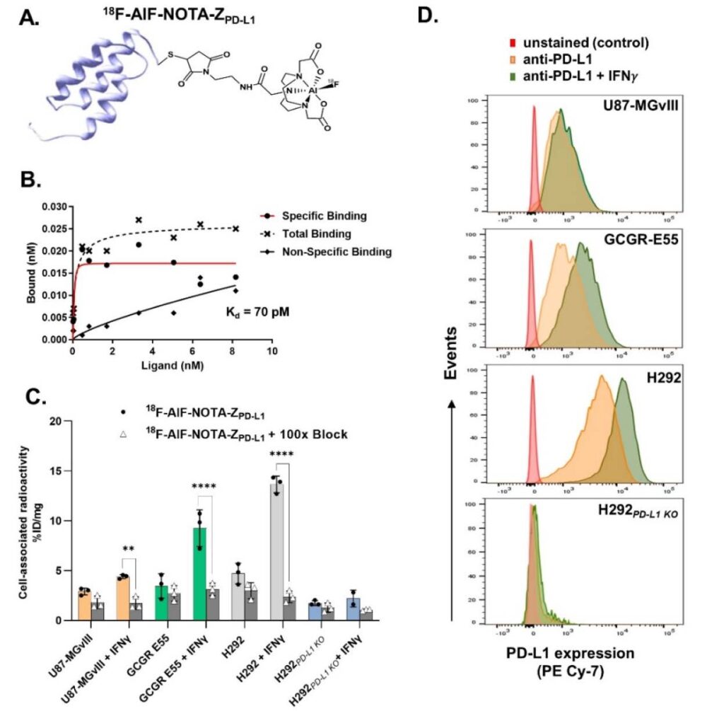
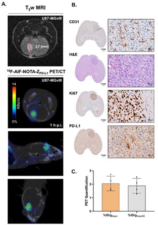
In this study, the research team led by Dr. Gabriela Kramer-Marek at The Institute of Cancer Research, London, demonstrated the suitability of the affibody molecule (ZPD-L1) radiolabeled with fluorine F-18 and gallium Ga-68 to noninvasively measure PD-L1 expression levels in orthotopic xenograft GBM models (Figure 1). Both 18F-AlF-NOTA-ZPD-L1 and 68Ga-NOTA-ZPD-L1 showed selective accumulation in PD-L1-positive tumors and detected GBM brain tumors with high contrast, corroborating T2-weighted MRI delineation of the tumors.
In this study, the use of our M-Series range of compact MRI was pivotal for quantifying tumor volume, monitoring growth and infiltrative phenotype following intracranial injection of human glioblastoma U87-MGvIII cells or Glioma GCGR-E55 Stem cells in mice, and provided a decision-making tool to plan the timing of the immuno-PET experiment. The Aspect Imaging M-series range offers cost-effective, low-maintenance, and compact MRI solutions for mice (M3), rats (M7), and non-human primates (M12), based on a unique permanent magnet design and an effective workflow supported by integrated physiological monitoring and a user-friendly operating system. The field strength of the M-Series (1 Tesla) is appropriate for rapid screening of tumor development, progression, and response assessment, making it optimal for lung imaging and the development of clinically translatable functional and/or molecular imaging approaches.
In summary, this study warrants further evaluation of anti-PD-L1 affibody-based immuno-PET as a potential biomarker for the non-invasive stratification of GBM patients and the optimization of PD-1/PD-L1 checkpoint blockade therapy