Imagine the world of medical research without the power of ultrasound technology. The journey of ultrasound began as early as 1790, with numerous theories and inventions shaping its course. The inception of the medical ultrasound technology that we’re familiar with today can be traced back to 1956, thanks to the collaborative efforts of Ian Donald, an obstetrician, and Tom Brown, an engineer. Over the years, a multitude of ultrasound variations have been conceived and produced, predominantly for diagnostic applications. We are delighted to draw attention to the impressive impact of high-frequency ultrasound (HFUS) in preclinical and research domains, despite its extensive use in clinical settings.
High-frequency ultrasound serves as an advanced diagnostic tool, utilizing sound waves that range from 20 to 30 MHz, and can extend up to 100 MHz in ultra-high-frequency ultrasound (UHFUS) scenarios. This leads to the creation of high-resolution images of targeted structures. The role of this technology has been pivotal in augmenting the precision and intricacy of imaging, thereby facilitating the identification and diagnosis of a variety of pathologies in preclinical research. It offers a range of advantages over other ultrasound options, including diverse applications.
The Impact of High-Frequency Ultrasound in Preclinical and Research Diagnostics
High-frequency ultrasound has revolutionized preclinical and research diagnostics by providing high-resolution images of superficial structures. Its non-invasive, safe, and versatile nature has made it an invaluable tool in various research applications, particularly in studies involving small animal models.
As technology continues to evolve, high-frequency ultrasound holds the potential for further advancements and applications in preclinical research, promising even greater precision and efficacy in data collection and analysis.
Enhanced Imaging Clarity
High-frequency ultrasound offers superior spatial resolution, allowing highly detailed images of subcutaneous body structures. By capturing images with greater clarity, HFUS enables researchers to identify abnormalities and pathologies more accurately in preclinical models.
Improved Diagnostic Accuracy
Numerous studies have indicated that high-frequency ultrasound significantly enhances diagnostic accuracy compared to relying solely on clinical examination. HFUS can detect the presence of tumors or metastases in many different tissues, monitor blood flow, assess systolic and diastolic function in the heart, and much more. As a result, the higher-resolution images provided by HFUS enable researchers to make more precise diagnoses, leading to more accurate research outcomes.
Non-Invasive and Safe
High-frequency ultrasound is a non-invasive and safe imaging technique. It does not involve exposure to ionizing radiation, making it a preferred choice over other imaging modalities. This non-invasiveness ensures minimal risk to testing subjects while delivering valuable diagnostic information in preclinical trials.
Real-Time Imaging
One of the significant advantages of ultrasound technology is its ability to provide real-time imaging. Researchers can observe the target area in motion, enabling them to assess and diagnose conditions more efficiently. Real-time imaging is particularly valuable during interventional procedures or when monitoring dynamic processes within the body.
Versatility in Research Applications
High-frequency ultrasound finds applications in various research settings due to its versatility. It has been successfully used in dermatology research to assess skin thickness, visualize skin structure, and monitor healing in acute and chronic wounds. Additionally, it has proven beneficial in ophthalmology research for examining the eye and its surrounding structures. Small animal imaging and musculoskeletal imaging are other areas where high-frequency ultrasound has demonstrated its versatility and utility.
High-Frequency Ultrasound Solutions From Scintica
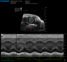
At Scintica, we offer innovative high-frequency ultrasound solutions for in vivo preclinical imaging in small animal models. Our Prospect T1 system is compact, cost-effective, and comes with a range of standard imaging modes to suit a wide variety of applications. It is primarily used in abdominal and anatomical imaging, cancer, cardiovascular and developmental biology, and ophthalmology and can also be used on models such as chick embryos, hamsters, and zebrafish.
Contact us today if your preclinical research applications could benefit from high-frequency ultrasound solutions.
References and further reading:
- Ai-Ho Liao, Pai-Chi Li,The Role of High Frequency Ultrasound in Multimodality Small Animal Imaging for Cancer Research, Journal of Medical Ultrasound, Volume 17, Issue 2, 2009, Pages 86-97, ISSN 0929-6441, https://doi.org/10.1016/S0929-6441(09)60115-6.