High-frequency ultrasound stands at the forefront of transformative technologies in preclinical research, offering unparalleled insights into the minutiae of biological processes. As researchers seek to unravel the complexities of disease and the impact of novel therapies, the precision and versatility of this modality have become indispensable. The ability to visualize the internal workings of small animals with exceptional clarity propels the boundaries of what can be discerned in vivo, marking a new epoch in diagnostic acumen.
Advanced Diagnostic Tool
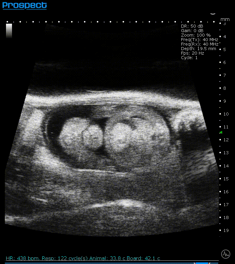
The utility of high-frequency ultrasound as an advanced diagnostic tool is underscored by its capacity to emit sound waves ranging from the standard 20 to 30 MHz to the more sophisticated 100 MHz in ultra-high-frequency systems. This spectrum allows for the acquisition of high-resolution images with up to 30 µm detail, a scale that brings cellular and subcellular structures into sharp relief. The real-time monitoring of hemodynamics and anatomical changes equips researchers with a dynamic view of physiological processes, enabling an unprecedented level of detail in disease characterization and therapeutic response assessment.
Non-invasive Imaging
The non-invasive nature of ultrasound imaging is a boon for preclinical studies. It permits serial measurements within the same subject, thus maintaining the integrity of the study while reducing the number of animals required. This aspect is not only ethically sound but also scientifically robust, as it allows for the tracking of disease progression or therapeutic efficacy over time with minimal physiological disruption.
Cost-effective
In the economic landscape of research, high-frequency ultrasound systems like the Prospect T1 emerge as cost-effective solutions. Their design is tailored for in vivo imaging in small animals, offering a compact and efficient alternative to larger, more expensive imaging setups. The integration of such systems within a laboratory setting circumvents the need for centralized imaging facilities, optimizing both time and financial resources.
Versatile Imaging Options
Versatility is another hallmark of high-frequency ultrasound systems. They support a plethora of imaging modalities including B-mode, M-mode, Color/Power Doppler, Pulsed Wave Doppler, Tissue Doppler, and Contrast imaging. Each modality serves a unique purpose, from evaluating cardiac function to assessing blood flow and visualizing the microvascular environment. This multifaceted capability ensures that high-frequency ultrasound can cater to a broad spectrum of preclinical research needs.
Interested in High-Frequency Ultrasound?
At Scintica, we recognize the critical role that high-frequency ultrasound plays in advancing preclinical research outcomes. Our commitment to innovation is reflected in the sophisticated imaging solutions we provide to the scientific community.
The Prospect T1 is our flagship, high-frequency ultrasound system designed for in vivo preclinical imaging in small animals such as mice and rats. It provides high-resolution images and advanced capabilities to monitor hemodynamic changes and observe anatomical structures in real time. The system offers access to the RAW digital RF data, enabling it to generate quantitative results and perform offline data analysis. Additional features include:
- A scanning platform equipped with a heated mouse or rat-sized platform and integrated physiological monitoring, making it easier to position the animal and acquire high-quality images.
- Various imaging options for various applications, including standard ultrasound capabilities such as B-mode, M-mode, Color/Power Doppler, Pulsed Wave Doppler, Tissue Doppler, and Contrast imaging.
- Ergonomically designed animal handling apparatus which ensures easy positioning and cleaning between imaging subjects. At the same time, the analytical tools and measurement packages help to speed up the process of generating results.
The Prospect T1 can be used within the laboratory and does not need to reside in a core facility. It is a cost-effective system that offers comprehensive measurement packages and can be used for various applications, including cardiovascular biology.
We invite researchers to explore the potential of high-frequency ultrasound to enhance their studies, confident in its ability to deliver non-invasive, cost-effective, and versatile imaging options. Discover more about how our high-frequency ultrasound systems can elevate your research. Join us in this journey of discovery, where every image brings us closer to the next scientific breakthrough.