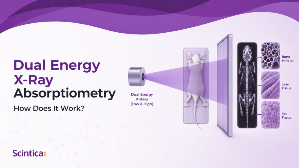
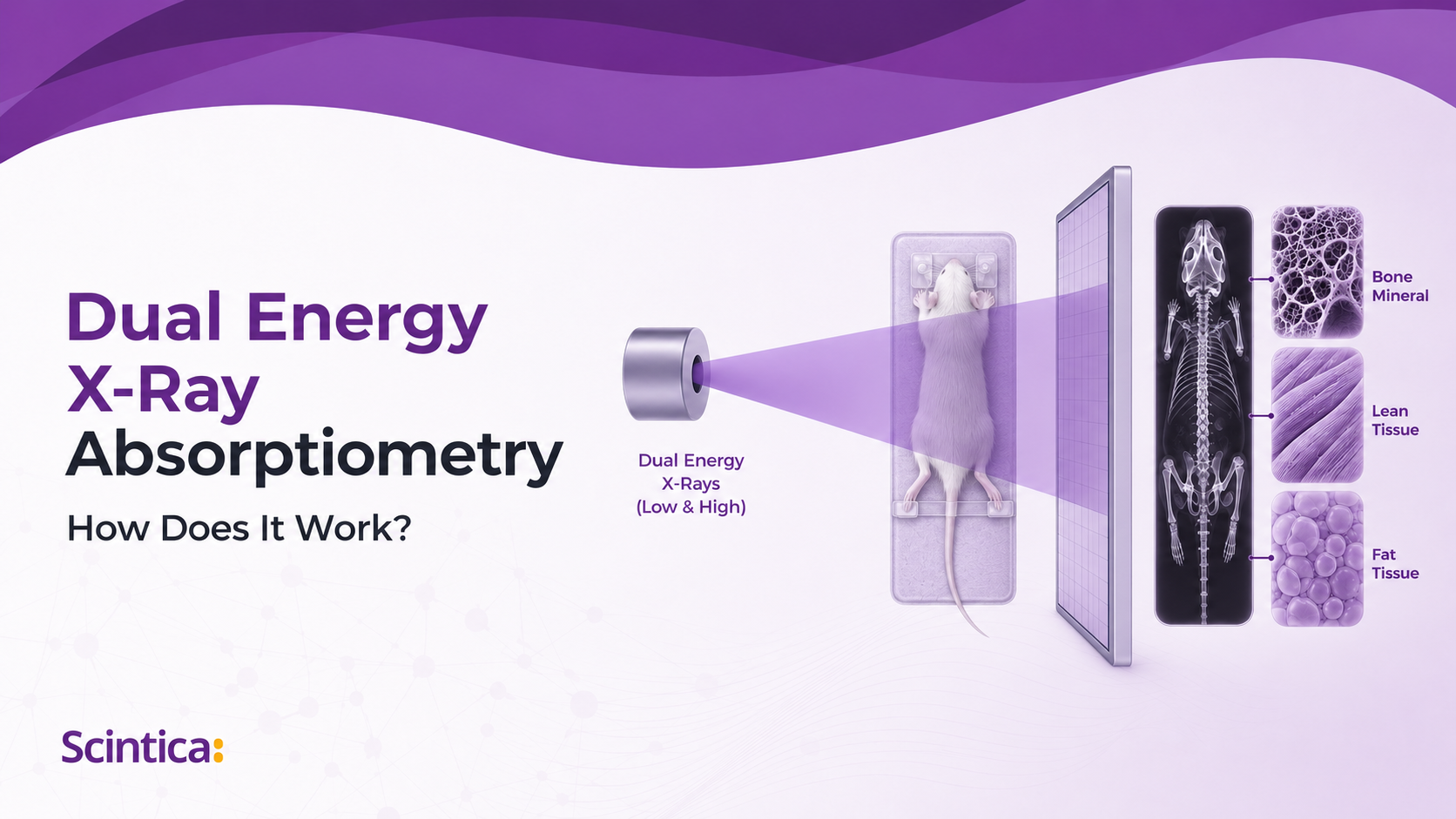
Dual Energy X-Ray Absorptiometry: How Does it Work?
Dual-energy X-ray absorptiometry (DXA) is a non-invasive imaging technique used to measure bone mineral density, bone mineral content, lean mass, and fat mass. In preclinical research, DXA allows researchers to assess body composition and skeletal changes over time, making it valuable for studies involving osteoporosis, obesity, metabolic disease, musculoskeletal disorders, arthritis, and drug safety.
Introduction
Dual Energy X-Ray Absorptiometry (DXA/DEXA) is a technique used to assess body composition, providing measurements of bone mineral density and content
along with lean and fat mass measurements. In essence, each pixel on the x-ray image is assigned to one of three categories – bone, fat, or lean mass.
Clinical DXA
DXA is considered the standard of care for diagnosing and following osteoporosis clinically, while the World Health Organization (WHO) considers it the gold standard in measuring bone mineral density, providing the most accurate results. Measurements are considered to be very precise and stable over time, and can allow for evaluation of structural, bone density, and body composition changes over time. Clinically, changes seen on traditional x-ray represent advanced disease which likely will not respond to treatment; changes can be observed much earlier using DXA, when interventions may be effective.
Preclinical DXA
In small-animal preclinical research, body composition and bone mineral density measurements are relevant across many research areas, including:
- Osteoporosis
- Rickets
- Osteomalacia
- Osteogenesis imperfecta
- Diabetes
- Metabolic syndrome
- Obesity
- Metabolic bone diseases and arthritis
- Bone and/or muscle regeneration
- Muscle-wasting diseases
These measurements can be used in arthritis, drug safety, and toxicology research to assess disease progression or regression in response to a therapeutic regimen or exposure to a compound of interest.
What Images and Data can be Generated Using a DXA System?
As will be described in more detail in the following section dual energy x-ray absorptiometry is the result of two x-ray images which are taken, one using low energy and one using high energy x-ray beams. These images are inherently co-registered as the animal is anesthetized so it does not move. The attenuation of the x-ray beams is measured at both energy levels and algorithms are used to determine which of the 3- compartment system each pixel falls into – fat mass, lean mass, or bone mineral content.
This acquisition results in three graphical outputs (1) x-ray attenuation image, (2) bone mineral density image, and (3) colorimetric image. Algorithms are used to generate the following measures:
- Bone mineral density (BMD) in g/cm²
- Bone mineral content (BMC) in g
- Bone area (BA) in cm²
- Tissue area in cm²
- Fat tissue as percentage and weight in % and g
- Lean tissue as percentage and weight in % and g
- Total weight in g
- Additionally, specific bone length measurements can be drawn on the 2D x-ray image
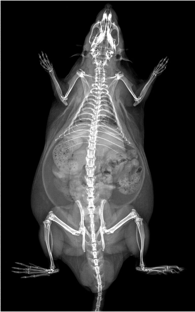
X-Ray attenuation

Bone Mineral Density

Colorimetric
How DXA Uses X-Ray Attenuation to Classify Tissue
Fundamentally, dual-energy X-ray absorptiometry (DXA) takes advantage of the attenuation of X-ray photons as they pass through the tissues of the body. The extent of attenuation varies with the energy of the photons and with the density and thickness of the material through which they pass.
How DXA Uses X-Ray Attenuation to Classify Tissue
For X-rays of the same energy, attenuation can be described using the following equation:
I = I0e−μM
I
Measured intensity of the X-ray
I0
Initial intensity of the X-ray beam
μ
Mass attenuation coefficient for the specific tissue, in cm²/g
M
Area density of the specific tissue, in g/cm²
At a given energy level, each tissue has a characteristic attenuation value. This constant is known as the mass attenuation coefficient.

Figure from Luo, Yunhua. 2017. Chapter 3 – Bone Imaging for Osteoporosis Assessment
Acquiring Low- and High-Energy Images
During DXA, two X-ray images are acquired: one using a low-energy beam and one using a high-energy beam. The X-rays are generated at the source, pass through the body, and reach the X-ray detector.
Because the animal does not move between acquisitions, the two images are inherently co-registered. Corresponding pixels therefore represent the same location in the animal and can be compared directly.
Classifying Bone Mineral and Soft Tissue
The first stage of the analysis determines whether each pixel should be classified as bone mineral or soft tissue. Bone mineral is a physically dense material composed primarily of phosphorus and calcium. These elements have relatively high atomic numbers and therefore produce substantial attenuation of the X-ray beams.
Soft tissue is a mixture of muscle, fat, skin, and water. It has a lower physical density and is primarily composed of elements such as hydrogen, carbon, and oxygen, which have lower atomic numbers and therefore produce less attenuation.
Bone Mineral
Soft Tissue
Primarily phosphorus and calcium
Muscle, fat, skin, and water
Higher physical density
Lower physical density
Relatively high atomic numbers
Primarily lower-atomic-number elements
Greater X-ray attenuation
Lower X-ray attenuation
At the same X-ray energy, bone mineral and soft tissue have different mass attenuation coefficients. The attenuation equation therefore becomes:

In this equation, the subscripts B and S refer to bone mineral and soft tissue, respectively.
Using Two Energies to Separate Bone and Soft Tissue
Because the mass attenuation coefficients differ at low and high X-ray energies, DXA produces two equations—one for each acquisition:
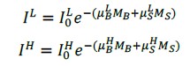
By comparing attenuation at the two energies, the system can determine which pixels represent bone mineral and which represent soft tissue.
Calculating Bone Area Density
The area density of bone mineral, MB, can be calculated using the following equation:
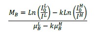
Where:

The ratio k is derived from image regions in which no bone is present; in these regions, MB equals 0. Once k has been determined, the equation can be solved for MB.
The MB value is calculated for each pixel to create the bone mineral density image. An edge-detection algorithm is then used to define the bone profile, from which bone area, bone mineral density, and bone mineral content can be calculated.
BA
Bone area (cm²)
BMD
Bone mineral density (g/cm²)
BMC
Bone mineral content (g)

Classifying Soft Tissue as Lean or Fat Mass
The equations above initially assume a two-compartment system: bone mineral or soft tissue. Any pixel that is not included in the bone area measurement is therefore classified as soft tissue.
The same set of equations can then be applied to the soft-tissue pixels to determine whether their attenuation is associated with lean or fat mass.
Because biological soft tissue inevitably contains molecules associated with both lean and fat tissue, each system must be calibrated using appropriate reference materials to assign soft tissue correctly. The values used depend on the lipid and non-lipid biological references, as well as the X-ray energies generated by the system.
Limitations of Two-Dimensional DXA
DXA produces two-dimensional images and cannot directly determine the thickness of the tissue through which the X-rays passed. Each pixel can therefore represent only one of the three tissue types in the three-compartment model:
Bone mineral · Fat mass · Lean mass
Soft tissue located over bone cannot be differentiated from the underlying bone within the same pixel. Likewise, a thick, diffuse bone cannot be directly compared with a thin, dense bone.
Visceral and subcutaneous adipose tissue that overlap within the trunk also cannot be differentiated. However, laterally located subcutaneous fat may occupy its own pixels and can therefore be differentiated from visceral fat.
Body Composition Analysis
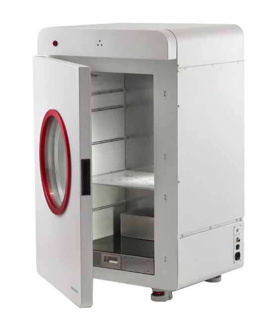
The iNSiGHT is a fully shielded X-ray cabinet which was specifically designed for preclinical small animal DXA applications, primarily using mice, rats, and similarly sized animals (10 to 500g). Measurements of body composition are taken quickly (~25 seconds), are non-invasive, and use very low-dose radiation; combined this makes the iNSiGHT DXA ideal for following the same animal over the course of a longitudinal study.
Measurements are made, using dual energy x-rays, to allow each pixel to be assigned to a 3-compartment model – that is either fat mass, non-bone/lean mass, or bone mineral content. DXA does not require any pre-treatment of the animal, contrast, or substrate injection, to acquire the data. It is also non-destructive and provides highly accurate and reproducible measures, and can be made on the whole animal or specific regions.
Data Analyzed by the iNSiGHT DXA
DXA analysis provides quantitative measurements of bone mineral density (BMD) in g/cm², bone mineral content (BMC) in g, bone area (BA) and tissue area in cm², and total weight in g. Fat and lean tissue are each reported as both a percentage of total tissue and a mass in grams. Specific bone-length measurements can also be obtained directly from the 2D X-ray image.