Yibo Quan ,1 Jie He ,1,2 Qi Zou,1,2 Liuxi Zhang,1,2 Qihui Sun, 2 Hongli Huang, 1 Wanglin Li, 1 Keping Xie, 2 Fang Wei1, 2
System Used:
IVIM System

Download Publication
ABSTRACT
Background: Immunotherapy, including adoptive cell therapy (ACT) and immune checkpoint inhibitors (ICIs), has a limited effect in most patients with colorectal cancer (CRC), and the efficacy is further limited in patients with liver metastasis. Lack of antitumor lymphocyte infiltration could be a major cause, and there remains an urgent need for more potent and safer therapies for CRC.
Methods: In this study, the antitumoral synergism of low molecular weight heparin (LMWH) combined with immunotherapy in the microsatellite stable (MSS) highly aggressive murine model of CRC was fully evaluated.
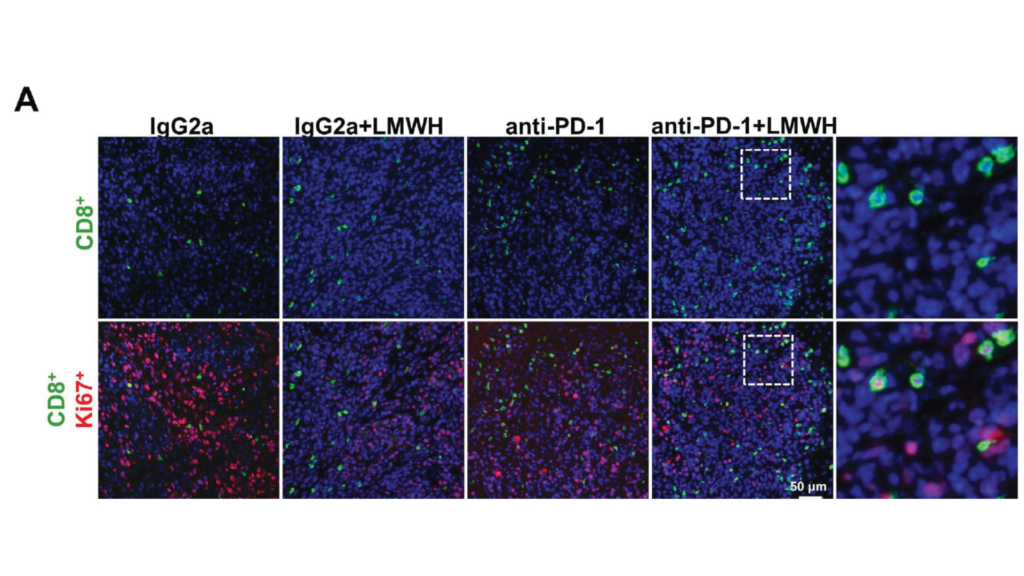
Results: Dual LMWH and ACT objectively mediated the stagnation of tumor growth and inhibition of liver metastasis, neither LMWH nor ACT alone had any antitumoral activity on them. The combination of LMWH and ACT obviously increased the infiltration of intratumor CD8+ T cells, as revealed by multiplex immunohistochemistry, purified CD8+ T-cell transfer assay, and IVIM in vivo imaging. Mechanistically, evaluation of changes in the tumor microenvironment revealed that LMWH improved tumor vascular normalization and facilitated the trafficking of activated CD8+ T cells into tumors. Similarly, LMWH combined with anti-programmed cell death protein 1 (PD-1) therapy provided superior antitumor activity as compared with the single PD-1 blockade in murine CT26 tumor models.
Conclusions: LMWH could enhance ACT and ICIs-based immunotherapy by increasing lymphocyte infiltration into tumors, especially cytotoxic CD8+ T cells. These results indicate that combining LMWH with an immunotherapy strategy presents a promising and safe approach for CRC treatment, especially in MSS tumors.
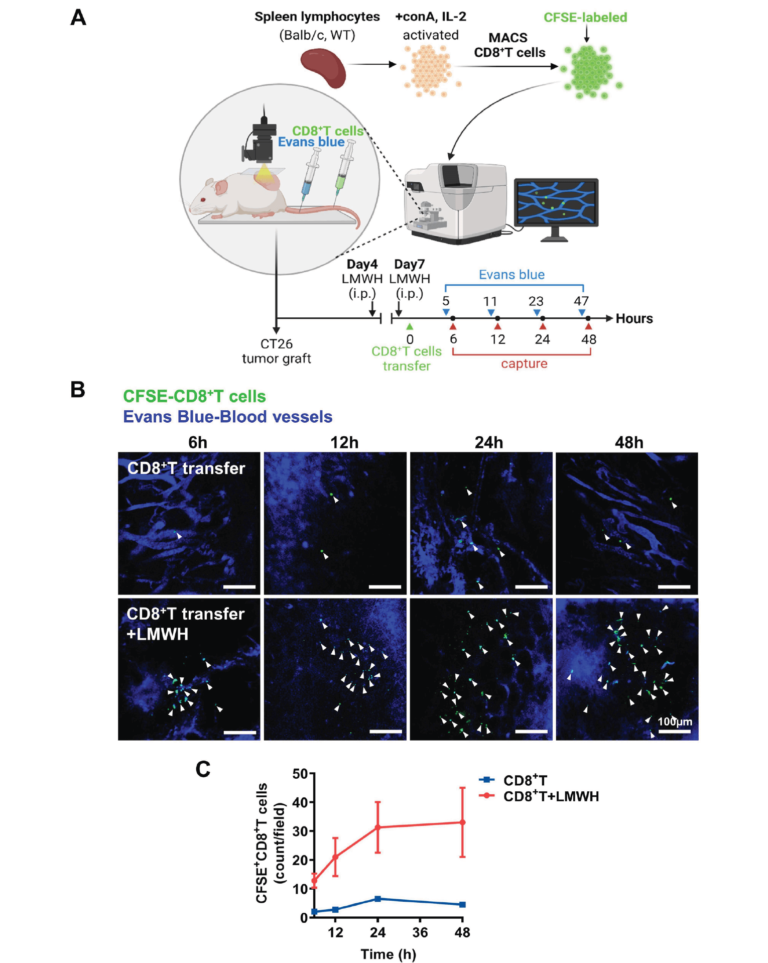
IVIM in vivo imaging of increased intratumor CD8+T-cell accumulation by LMWH treatment. (A) Flow chart of CFSE-CD8+ T-cell infiltration observed by IVIM in vivo imaging. (B) Representative images of CFSE-CD8+ T-cell infiltration were taken by IVIM in vivo imaging at 6 hours, 12 hours, 24 hours, and 48 hours after cell transfer (blue, angiography Evans blue; green, CFSE-CD8+ T cells). (C) Quantification of CFSE-CD8+ T cells migrating to tumors through blood vessels in live images. conA, concanavalin A; IL, interleukin; CFSE, carboxyfluorescein diacetate succinimidyl ester; i.p., intraperitoneally; LMWH, low molecular weight heparin; WT, wild type.