(July 29, 2020) WEBINAR: Cancer Research in Small Animals: A Review of Recent Publications Using High Frequency Ultrasound
This webinar hosted by Scintica Instrumentation discussed the benefits of ultrasound imaging in cancer research. Ms. Tonya Coulthard was the presenter of this webinar, she touched on several topics throughout the presentation as they pertain to cancer biology and preclinical imaging. First off a brief overview of the different types of tumor models, followed by a look at the differences between optical and ultrasound imaging in cancer research, and finally a brief thought on image guided injections for placement of tumor cells in various models.
During the last half of the webinar Tonya walked participants through some of the recent publications using high frequency ultrasound imaging to monitor tumor growth, study therapeutic effect on progression, as well as some publications using a variety of contrast agents. The work highlighted researchers using the Prospect T1 system manufactured by S-Sharp.
Ultrasound imaging is a non-invasive imaging technique using sound waves to produce images of a variety of internal structures, including tumors. Ultrasound is used clinically, as well as preclinically to detect and assess tumor location and size, as well as therapeutic response. Many tumor models use mice and/or rats, necessitating high frequency ultrasound with image resolution down to 30µm.
Topics discussed in this webinar included:
- Different types of tumor models
- Ultrasound vs. optical imaging in cancer research
- Image guided injections of cancer cells
- Publication overview

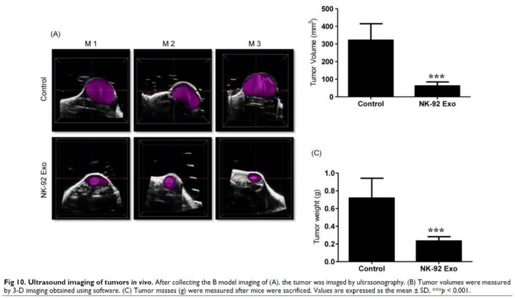
The figure above is from one of the papers that Tonya reviewed during the webinar (Zhu et al. Theranostics. 2017; 7(10): 2732-2745.)
- Questions
- Slide Deck

About the Speaker (s)

Tonya Coulthard, MSc
Manager, Imaging Division
Scintica, London, Ontario
Tonya Coulthard holds an MSc from the University of British Columbia in Experimental Medicine. Throughout her academic training she focused on a variety of diseases ranging from prion to chronic obstructive pulmonary diseases. In her professional career she has worked with several imaging and medical device companies; in these roles she has interacted with researchers around the world covering a very diverse range of research applications. In her present role at Scintica Instrumentation Tonya manages our Imaging Division; in this role she works in supporting our customers in understanding the products offered and how these instruments help to meet their research needs.