A Bone Morphogenetic Protein-2 (BMP-2) Load Scaffold for Recruiting and Harvesting of Stem- and Immune- Cells for Therapy:
Focus on the Role of the iNSiGHT Dual Energy X-Ray Absorptiometry (D(E)XA) System in the Development and Optimization of Osteo-Organoids
System Used:
iNSiGHT
In their recent study published in Science Advances, Dai and co-workers demonstrated how a BMP-2 osteo-organoid-loaded scaffold implanted into the muscle pocket near the femur of a mouse can be used as a niche to support the abundant recruitment of stem cells and mature immune cells. This in vivo osteo-organoid consisting of a BMP-2-loaded gelatin scaffold supported the production of hematopoietic stem/progenitor cells (HSPCs), mesenchymal stem cells (MSCs), lymphocytes, and myeloid, via a three-stage process: fibroproliferation, osteochondral differentiation, and marrow generation. Osteochondral-stage and marrow-stage cells were successfully harvested and used to treat hepatic injuries and to reconstitute the impaired immune system in a radiation-damage assay, respectively. These results demonstrate the potential of the BMP-2 osteo-organoid as a sustainable source of high-quality stem- and immune- cells for cells therapies and regenerative medicine applications.
In this study, the authors exploited the unique ability of the iNSiGHT dual energy X-ray absorptiometry (D(E)XA) system to rapidly and non-invasively monitor changes in bone mineral density (BMD) and evaluate the time- and dose- dependence of the successful osteo-organoid formation following the implantation of the BMP-2 loaded gelatin scaffold (Figure 1). The authors used the iNSiGHT D(E)XA system to demonstrate that the development of the osteo-organoid did not significantly alter the overall body and femur BMD, and to confirm the successful increase in BMD in the excised osteo-organoid, allowing them to rapidly find the optimal and cost-effective dose for osteo-organoid construction in further experiments.
New features of the INSIGHT software allow the evaluation of BMD, fat, or lean mass in specific regions of interest (ROIs) during post-processing, thereby warranting further evaluation of the dataset generated in this study, especially the comparison between the BMD of the osteo-organoid ROI acquired in vivo with the BMD, weight, and histopathological scoring of the excised osteo-organoid. This data would help calibrate and further validate the iNSiGHT DEXA system, and by extension clinical D(E)XA imaging, as a pivotal tools to monitor non-invasively osteo-organoid formation in vivo to further accelerate the clinical translation and evaluation of this promising approach for harvesting stem cells and immune cells for therapeutic purpose.
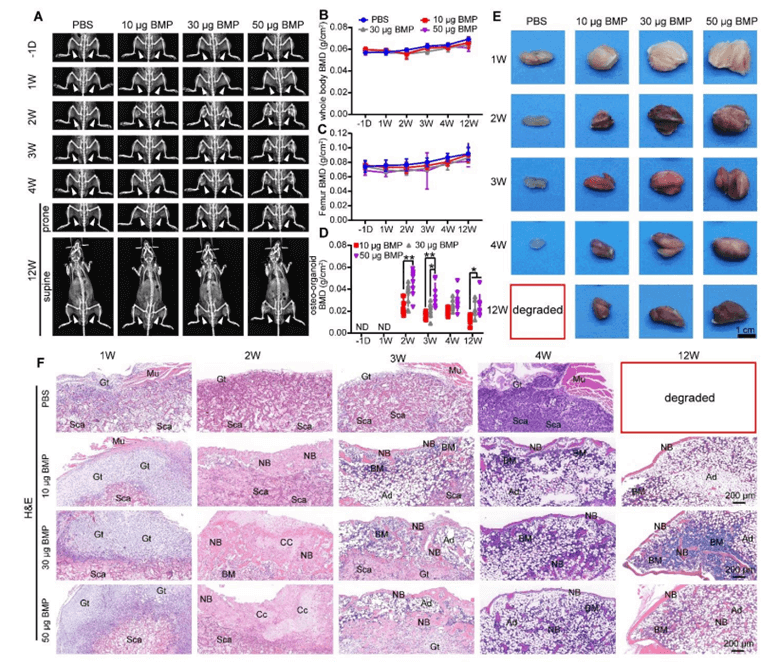
Figure 1. A) Dual-energy X-ray absorptiometry ((D(E)XA)) images of mice injected with varying dosages (10, 30, or 50 µg) of rhBMP-2 imaged 6 times up to 12 weeks. B, C, D) Bone mineral density (BMD) analysis for whole body, femur, and the osteo-organoid for the varying dosages and time points (week 1,2,3,4, and 12 post-implantations. Obvious bone generation was observed after only 2 weeks when comparing to the PBS control group, as confirmed by E) visual inspection and F) hematoxylin and eosin (H&E) staining of the excised tissue, highlighting granulated tissue (Gt), scaffolds (Sca), chondrocytes (Cc), muscle (Mu), bone marrow (BM), new bone (NB), and adipocytes (Ad). D(E)XA imaging confirmed a dosage of 30 µg as an adequate and cost-effective dose for the development of osteo-organoids for further experiments.
Author
Tyle Lalonde Ph.D. – Product Manager – Scintica:
References
Dai, Kai, et al. “A BMP-2–triggered in vivo osteo-organoid for cell therapy.” Science Advances 9.1 (2023): eadd1541.