Blood-pool MRI assessment of myocardial microvascular reactivity
System Used:
OxyLite
Authors:
Sadi Loai, Beiping Qiang, Michael A. Laflamme, Hai-Ling Margaret Cheng
Abstract:
Purpose: The ability to non-invasively image myocardial microvascular dilation and constriction is essential to assessing intact function and dysfunction. Yet, conventional measurements based on blood oxygenation are not specific to changes in blood volume. The purpose of this study was to extend to the heart a blood-pool MRI approach for assessing vasomodulation in the presence of blood gas changes and investigate if sex-related differences exist.
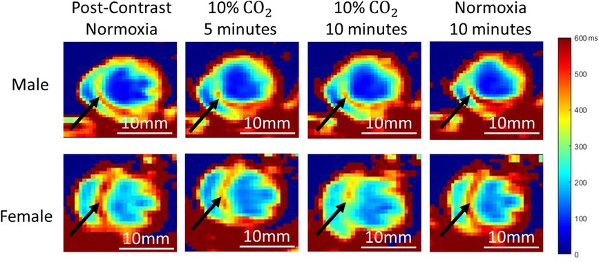
Methods: Animals [five male and five female healthy Sprague Dawley rats (200–500 g)] were intubated, ventilated, and cycled through room air (normoxia) and hypercapnia (10% CO2) in 10-minute cycles after i.v. injection of blood-pool agent Ablavar (0.3 mmol/kg). Pre-contrast T1 maps and T1-weighted 3D CINE were acquired on a 3 Tesla preclinical MRI scanner, followed by repeated 3D CINE every 5 min until the end of the gas regime. Invasive laser Doppler flowmetry of myocardial perfusion was performed to corroborate MRI results.
Results: Myocardial microvascular dilation to hypercapnia and constriction to normoxia were readily visualized on T1 maps. Over 10 min of hypercapnia, female myocardial T1 reduced by 20% (vasodilation), while no significant change was observed in the male myocardium. After return to normoxia, myocardial T1 increased (vasoconstriction) in both sexes (18% in females and 16% in males). Laser Doppler perfusion measurements confirmed vasomodulatory responses observed on MRI.
Conclusion: Blood-pool MRI is sensitive and specific to vasomodulation in the myocardial microcirculation. Sex-related differences exist in the healthy myocardium in response to mild hypercapnic stimuli.